Download
1 / 37
460 likes | 963 Vues
HISTOLOGY & HISTOCHEMISTRY By Professor Abdel-Majeed Safer. Chapter 1. The branch of science that deals with the chemical composition of the cells and tissues of the body. What is HISTOLOGY ?.

E N D
HISTOLOGY & HISTOCHEMISTRY By Professor Abdel-Majeed Safer Professor Abdel-Majeed Safer Histology & Histochemistry
Chapter 1 Professor Abdel-Majeed Safer Histology & Histochemistry
The branch of science that deals with the chemical composition of the cells and tissues of the body. What is HISTOLOGY ? Histologyis the study of the tissue of the body and how these tissues are arranged to constitute organs. Histology involves all aspects of tissue biology with the focus on how cells’ structure and arrangement optimize functions specific to each organs. Histochemistry & Cytochemistryis the study of the chemical composition of the tissue and cell of the body. Or precisely, the microscopic study of the chemical characteristics of tissues and cells, through the use of substances (dyes etc.) producing identifying chemical reactions. Professor Abdel-Majeed Safer Histology &Histochemistry
HISTOLOGY AND HOW IT IS STUDIED Professor Abdel-Majeed Safer Histology & Histochemistry
HISTOLOGY and Its Method of Study Preparation of Tissues for Study Fixation Embedding & Sectioning Light Microscopy Bright field microscopy Fluorescence microscopy Phase-contrast microscopy Interference microscopy Confocal microscopy Polarizing microscopy Electron Microscopy Transmission Electron Microscopy TEM Scanning Electron Microscopy SEM Professor Abdel-Majeed Safer Histology & Histochemistry
HISTOLOGY and Its Method of Study Autoradiography Cell & Tissue culture Histochemistry & Cytochemistry Detection Methods using specific Interactions between molecules Immunohistochemistry Hybridization Techniques Problems in the study of Tissue Sections Professor Abdel-Majeed Safer Histology & Histochemistry
HISTOLOGY and Its Method of Study Histology is the study of the tissue of the body and how these tissues are arranged to constitute organs. Histology involves all aspects of tissue biology with the focus on how cells’ structure and arrangement optimize functions specific to each organs. Tissues are made of two interacting components: cells and extracellular matrix. The ECM consists of many kinds of molecules, such as collagen fibrils. there is, thus, an intense interaction between cells and matrix, with many components of the matrix . Most organs are formed by an orderly combination of several tissues, except the CNS, which is formed almost solely by nervous tissue. The precise combination of these tissues allows the functioning of each organ and of the organism as a whole. The small size of cells and matrix components makes histology depedent on the use of microscope. Professor Abdel-Majeed Safer Histology & Histochemistry
HISTOLOGY and Its Method of Study Preparation of Tissues for Study Fixation see Fig 1 Is to avoid tissue digestion by enzymes within the cells (autolysis) or by bateria and to preserve structure and molecular composition. Chemical methods - the tissues are immersed in solution of stabilizing or cross-linking agents called fixatives. or less frequently Physical methods (deep freeze). In routine LM, formalin ; a buffered isotonic solution of 37% formaldehyde. Formaldehyde and glutaraldehyde are commonly used and known to react with amine groups NH2- of tissue proteins. Glutaraldehyde, is reinforced by virtue of being a dialdehyde which croos-links proteins. In EM, where high resolution is require, a buffered glutaraldehyde followed by a second fixation in buffered OsO4 for fine structural studies. OsO4 is used to preserve and stain lipids and proteins. Professor Abdel-Majeed Safer Histology & Histochemistry
HISTOLOGY and Its Method of Study Dehydration To extract the water from the tissue, through graded series of alcohol (70% - 100% ethanol). Clearing Ethanol is then replaced by a clearing agent that is miscible with both alcohol and the embedding medium. E.g. xylene Paraffin Melted paraffin 52- 60oC in an oven. Embedding and Sectioning Tissues are embedded in a solid medium to facilitate sectioning. To obtain thin sections with the microtomes, tissues must be infiltrated after fixation with embedding solutions that impart a rigid consistency to the tissue. Such as paraffin and plastic resins. Professor Abdel-Majeed Safer Histology & Histochemistry
HISTOLOGY and Its Method of Study Tissues to be embedded in plastic resin are also dehydrated in ethanol, cleared and embedded in plastic solvent, which are then hardened by means of cross-linking polymerizes. Sectioning and Microtomysee Fig.2 The hard blocks containing the tissues are placed in microtome. Cut 1-10 um (ium= 1/1000 mm. 1nm = 0.001um = 10-6 mm. 1A = 0.1nm 10-4um. Sections are are floated on water and then transferred to glass slides to be stained . Frozen sections are also performed by rapid freezing (physical , not chemical fixation), using a freezing mictotome or cryostat. This is mainly for rapid invistigation in hospitals and for histochemical studies, as enzymes and small molecules and lipids are very sensitive. Professor Abdel-Majeed Safer Histology & Histochemistry
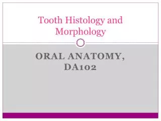
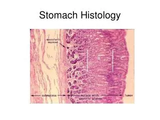
Columnar epithelium lining the small intestine • Micrograph stained with hematoxylin and eosin (H&E). Tissue stained with the periodic acid-Schiff (PAS) reaction for glycoproteins. Tissue stained with hematoxylin and eosin (H&E).
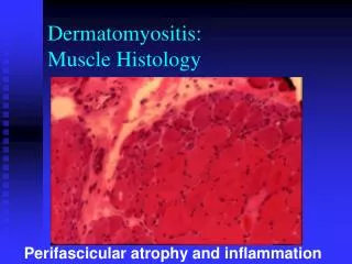
HISTOLOGY and Its Method of Study Histological Stains For routine histological work it is customary to use two dyes, one stains certain components, and the other stains different ones (counterstain). Toluidine blue Basophilic and Acidophilic Staining Basophilic substance is the one that takes up basic stain, acidophilic is the one that takes up an acidic stain. Hematoxylin is a basic stain or cationic stain, while Eosin is an acidic stain or anionic stain. What do the color seen in Hematoxylin sections indicate? Ordinary stains such H&E provide only general information about the chemical composition of the components that they color. Professor Abdel-Majeed Safer Histology & Histochemistry
Light Microscopy Bright field microscopy Stained slides are examined by means of ordinary light that passes through the specimen. See Fig.1-3. for details. Flourescence Microscopy When certain substances are irradiated by light of a proper wavelength, they emit light with a longer wavelength. This phenomenon is called Flourescence. In Flourescence Microscopy, tissue sections are irradiated with ultraviolet UV light and the emission is in the visible portion of the spectrum. The fluorecent substances appear brilliant on a dark background. Thus, the microscope has a strong UV light source and special filters that select rays of different wavelengths by the substances. Fluorescent compounds with affinity for specific cell macromolecules may be used as Fluorescent stains. E.g. Acridine Orange (bind to DNA and RNA).See Fig.1-4a e.g..Hoechst stain and DAP1 specifically bind to DNA giving a blue flourecence under UV . Professor Abdel-Majeed Safer Histology & Histochemistry
Fluorescent Microscopy Culture of kidney cells DAPI (4’,6-diamino-2-phenylindole) Kidney cells acridineorange
Phase-contrast microscopy and Interference microscopy Unstaned biological specimens are usually transparent and difficult to view in detail, beacause all parts of the specimen have the same optical density. Phase-contrast microscopy uses a lense system that produces visible images from transparent objects (Fig. 1-5). Principle: Light changes its speed when passing through cellular and extracellular structures with different refractive indices. Thses changes are used by Phase-contrast system to cause the strctures to appear lighter or darker in relation ro each other. So, living cells and tissue cultures can be observed ( no need for fixation or staining). Phase-contrast microscope is essential tool in tissue culture labs. also, differential Interference microscopy, which produces images with a more apparent 3-D aspect than routine Phase-contrast microscopy (Fig. 1-5). Professor Abdel-Majeed Safer Histology & Histochemistry
Bright field microscopy Interference microscopy Phase contrast microscopy
Confocal microscopy • With a regular bright-field microscopy the beam of light is relatively large and fills the specimen. • Stray light reduces contrast within the image; confocal microscopy avoids stray light and achieves greater resolution by using: • A small point of high intensity light providing by a laser . • A plate with a pinhole aperture infront of the image detector. • the point light source, the focal point of the lens, and the detector’s pinpoint aperture are all optically conjugated or alighned to each other in the focal plane (CONFOCAL) and unfocused light does not pass through the pinhole. This greatly improves resolution of the object in focus and allows the localization of specimen components with much greater precision than with bright-filed microscope. Fig. 1-6. Professor Abdel-Majeed Safer Histology & Histochemistry
Figure 1–6. • Principle of confocal microscopy. Although a very small spot of light originating from one plane of the section crosses the pinhole and reaches the detector, rays originating from other planes are blocked by the blind. Thus, only one very thin plane of the specimen is focused at a time. The diagram shows the practical arrangement of a confocal microscope. Light from a laser source hits the specimen and is reflected. A beam splitter directs the reflected light to a pinhole and a detector. Light from components of the specimen that are above or below the focused plane is blocked by the blind. The laser scans the specimen so that a larger area of the specimen can be observed.
Polarizing microscopy Allows the recognitio, in of structures made of highly organized molecules. When normal light passes through a polarizing filter, it exists vibrating in only one direction. If a second filter is placed in the microscope above the first one, with its main axis perpendicular to the first filter, no light passes through. If, tissue structures containing oriented macromolecules are located between the two polarizing filters, their repetitive structure rotates the axis of the light emerging from the polarizer and they appear as bright structures against a dark background Fig.1-7. So, crystalline substances or substances containing highly oriented molecules such as cellulose, collagen, micromolecules and microfilaments have the property of birefringes. Professor Abdel-Majeed Safer Histology & Histochemistry
bright-field and polarizing microscopy Bright-field microscopy Plarizingmicroscopy
Figure 1–7. • Tissue appearance with bright-field and polarizing microscopy. Polarizing light microscopy produces an image only of material having repetitive, periodic macromolecular structure; features without such structure are not seen. Shown here is a piece of thin mesentery that was stained with red picrosirius, orcein, and hematoxylin, and was then placed directly on a slide and observed by bright-field and polarizing microscopy. (a): Under routine bright-field microscopy collagen fibers appear red, along with thin dark elastic fibers and cell nuclei. (b): Under polarizing light microscopy, only collagen fibers are visible and these exhibit intense birefringence and appear bright red or yellow; elastic fibers and nuclei lack oriented macromolecular structure and are not visible.
Electron microscopy TEM and SEM are based on the interaction of electrons and tissue components. The wavelength in electron beam is much shorter than of light. Allowing a thousand-fold increase in resolution. Transmission Electron Microscopy TEM with a resolution of 0.3nm (Fig.1-8a). Which allows magnification of up to 800,000 times to be viewed with details. (1nm = 0.001um = 10-6 mm. 1A = 0.1nm 10-4um). Principle: A beam of electrons can be deflected by electromagnitic fields. E beam is produced by a cathode and passes down the column in a vacuum. The beam can be focused by means of electric coils (electromagnetic lenses). First lens is Condenser lens, which focus beam on the specimen section. Second lens is Objective lens, when electrons pass the specimen without interacting with it, it reaches the Objective lens. This form a focused, magnified image, and reach a viewing screen. The image is white and black and gray. (bright areas indicate electron passing through, darker areas – electron dense- indicate electrons were absorbed or deflected). Professor Abdel-Majeed Safer Histology & Histochemistry
TEM requires very thin sections (40-90 nm), so plastic embedding blocks are cut with glass or diamond knives. Sections are collected on small copper grids and transferred into the microscope to be analyzed. Freeze-fractyre, cryofracture, freeze etched combined with TEM have been useful for examining membrane structure. Scanning Electron Microscopy SEM It permits 3-D views of the surfaces of cells, tissues, and organs. Beam does not pass through the specimens (Fig.1-8a). The sample is dried and coated with a very thin layer of metal atoms through which electrons do not pass readily. When the beam is scanned from point to point across the specimen it interact with the metal atoms and produces reflected electrons or secondary electrons emitted from the metal. These are captured by a detedtor and the resulting signal is processed to produce a black-and-white image on a monitor (Fig.1-8b). Professor Abdel-Majeed Safer Histology & Histochemistry
Autoradiography Is a method of localizing newly synthesized macromolecules (DNA, RNA, protein, glycoproteins, and polysaccharides) in cells or in tissue sections. Radioactively labled metabolites (nucleotides, amino acids) incorporated into the macromolecules emit weak radiation that is restricted to the cellular regions where the molecules are located. Radiolabel cells or mounted tissue sections are coated in a darkroom with photographic emulsion containing silver bromide crystals, which acts as microdetectors of this radiation in the same way that they respond to light in common photographic film. After some times, the slides are developed photographically. The silver bromide crystals reduced by the radiation are reduced to small black grains of metallic silver, indicating location of radiolabled macromolecules in the tissue. This tech can be used for both LM and EM (Fig.1-9). Practically, it is possible to know which cells is a tissue produce more protein and which cell produce less, because the number of silver grains formed over the cells is proportional to the intensity of protein synthesis. Professor Abdel-Majeed Safer Histology & Histochemistry
Autoradiographs are tissue preparations in which particles called silver grains indicate the regions of cells in which specific macromolecules were synthesized just prior to fixation Black “silver grains” are visible over regions with secretory granules and the duct indicating glycoprotein locations. X1500.
Autoradiographs are tissue preparations in which particles called silver grains indicate the regions of cells in which specific macromolecules were synthesized just prior to fixation The same tissue prepared for TEM autoradiography shows silver grains with a coiled or amorphous appearance again localized mainly over the granules (G) and in the gland lumen (L). X7500.
Cell & Tissue Culture • Live cells and tissues can be maintained and studied outside the body. cell culture has been very useful in isolating the effects of single molecules on specific types of cells. It also allows the direct observation of the behavior of living cells under the phase contrast microscope. Many experiments that cannot be performed in the living animal can be accomplished in vitro. • The cells and tissues are grown in complex solutions of known composition (salts, amino acids, vitamins) to which serum components or specific growth factors are added. • Cells must be dispersed mechanically or enzymatically. • The cells are then cultivated in a clear dish to which they adhere, as single-layer cellss(Fig.1-5). This is called primary cell culture, which makes cell line. Normal or pathologic tissue have been maintained in vitro ever since they have immortalized and now constitute a permanent cell line. • Cell culture has been widely used for study of the metabolism of normal and cancerous cells and for the development of new drugs. Professor Abdel-Majeed Safer Histology & Histochemistry
Histochemistry & Cytochemistry They are methods for localizing cellular structures in tissues using enzyme activity present in these structures. Usually it is applied to unfixed or mildly fixed tissue, sectioned on a cryostat or freezing microtome to avoid adverse effects of heat, paraffin and chemicals on enzymatic activity. 1. Tissue sections are immersed in a solution containing the substrate of the enzyme to be localized. 2. The enzyme is allowed to work on the substrate. 3. The section is put in contact with the marker compound. 4. This compound reacts with a molecule produced by enzymatic action on the substrate. 5. The final reaction product (insoluble and visible in LM or EM) only if it is colored of electron- dense, precipitate over the site that contains the enzyme. e.g. Phosphatases, split the bond between a PO4 group and an alcohol residue of phosphorylated molecules. The visible insoluble reaction product of phosphateses is lead phosphate or lead sulphide. At alkaline pH and cid phosphatese. (Fig.1-10). Professor Abdel-Majeed Safer Histology & Histochemistry
Dehydrogenases Remove H from one substrate and transfer it to another. Mitochondria can be specifically identified by this method, dehydrogenases are key enzymes in the citric acid cycle. Peroxidases Present in several types of cells, promotes the oxidation of certain substates with the transfer of H ions to H2O2, forming H2O. Fixed sections are incubated in a solution of H2O2 and 3,3”-diamino-azobenzinedine DAB. DAB is oxidized in the presence of peroxidaze, resulting in an insoluble , brown, electron-dense ppt w permit the localization of peroxidase activity by LM and EM. Professor Abdel-Majeed Safer Histology & Histochemistry
Artifacts Kind of flaw that is due to faulty technique: Postmortem degeneration Shrinkage Precipitants Wrinkles and Folds Nicks in the microtome knife Rough Handling Problems in the study of Tissue Sections (Fig.1-15) Professor Abdel-Majeed Safer Histology & Histochemistry
END of Chapter 1 Professor Abdel-Majeed Safer Histology & Histochemistry