Download
1 / 1
20 likes | 166 Vues
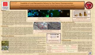
A. B. 96 kDa. 43 kDa. M ALLERY LAB. Presentation # 3123 Abstract # 118623. Feasibility of Local Nanoparticle Delivery for Oral Cancer Chemoprevention. Andrew S. Holpuch 1 , Garrett J. Hummel 1 , Meng Tong 1 , Ping Pei 1 , Ping Ma 2, Russell J. Mumper 2 , and Susan R. Mallery 1.

E N D
A B 96 kDa 43 kDa MALLERY LAB Presentation # 3123 Abstract # 118623 Feasibility of Local Nanoparticle Delivery for Oral Cancer Chemoprevention Andrew S. Holpuch1, Garrett J. Hummel1, Meng Tong1, Ping Pei1, Ping Ma2, Russell J. Mumper2, and Susan R. Mallery1 1Department of Oral and Maxillofacial Surgery and Pathology, College of Dentistry, The Ohio State University, Columbus, OH; 2Division of Molecular Pharmaceutics, School of Pharmacy, University of North Carolina at Chapel Hill, Chapel Hill, NC C D B A Abstract: Localized drug delivery provides a major pharmacologic advantage by obtaining therapeutically effective local levels without inducing systemic effects. Local delivery to the oral cavity often requires the use of aqueous gels, such as the formulation recently used by our labs in a Phase I/II chemoprevention trial. Notably, some optimal chemopreventive compounds, such as vitamin A derivatives, are not stable in aqueous formulations. Objective: This study investigated whether nanoparticles are a viable strategy for delivery of lipophilic chemopreventive compounds to human oral keratinocytes and human oral mucosal explants. Methods: These studies entail a series of in vitro assays that employ model nanoparticle compounds which were selected on the basis of composition, size and capacity for monitoring localization via fluorescence. Results to date reflect nanoparticle delivery to in vitro monolayer-cultured, characterized OSCC cell lines. Data were analyzed using the Student’s two-tailed t-test. RESULTS: Overall, nanoparticle-probe (NanoP) delivery resulted in higher final intracellular fluorescence levels relative to naked-probe (NakedP). Internalized fluorescence data show delivery method, time and probe concentration affect intracellular levels. Short time-course (1-hour) results demonstrate higher levels following NakedP administration [1000nM: average NakedP internalization (21.08±5.33) greater than NanoP internalization (12.87±4.32) p=.0025, n=18]. In contrast, longer duration (6-hour), higher probe concentrations demonstrated average NanoP internalizations (2500nM: 43.53±8.42, 5000nM: 64.44±12.71) that were significantly higher than NakedP internalizations (2500nM: C Figure 2. Nanoparticles associated with HNSCC cells in monolayer. A. SCC9, 30 min., 2500nM IDA-NP (400x) B. SCC15, 6 hr., 1µg BODIPY FL-C12 (400x) C. SCC15, 6 hr., 1µg BODIPY FL-C12 (600x) D. SCC15, 1 hr., 15nM QDot (600x) The extent of nanoparticle internalization/association is clearly seen in the perinuclear vesicles (A,B,C: green; D: red). Exogenous fluorescence was quenched in Figure 2, B and C. The mechanism of nanoparticle uptake in HNSCC cells is unknown, but studies are underway to determine the mechanism of endocytosis. Figure 4. Idarubicin-nanoparticle (NanoP) vs. Free-Idarubicin Internalization (NakedP) The internalization of NakedP was more rapid than NanoP after 1 hour at 1000nM (NakedP: 21.08 ± 5.33 vs. NanoP: 12.87 ± 4.32). However, the maximum uptake of NanoP was greater than NakedP after 6 hours at 2500nM (NanoP: 43.53 ± 8.42 vs. NakedP: 27.61 ± 5.66) and 5000nM (NanoP: 64.44 ± 12.71 vs. NakedP: 31.06 ± 7.24). Results and Discussion: 27.61±5.66, p=.0002, n=18; 5000nM: 31.06±7.24, p=.0001, n=18]. Additional studies which utilized extracellular quenching agents following introduction of BODIPY FL-C12-nanoparticles established OSCC nanoparticle internalization. CONCLUSIONS: Our data confirm the feasibility of nanoparticle-based compound delivery to oral keratinocytes and support the premise that nanoparticle delivery achieves higher sustained intracellular levels relative to bolus administration. Studies to evaluate the extent of nanoparticle penetration in intact oral mucosal tissues and mechanisms of nanoparticle internalization are ongoing. [NIH R01CA95901, R21CA132138 and T32DE14320.] Idarubicin Nanoparticle vs. Free-Idarubicin Internalization. The initial treatment of SCC9 cells with IDA-NPs in LabTek chamber slides suggested that the nanoparticles were internalized by the cells in monolayer, as seen in the photomicrograph in Figure 2a. Further, the quantitative comparison of IDA-NP (NanoP) versus free-IDA (NakedP) internalization shows an accelerated uptake of free-IDA compared to IDA-NP, but the overall maximum amount of uptake is higher for IDA-NP. Interestingly, the free IDA showed relatively constant levels of uptake, while the IDA-NP portrayed a dose- and time-dependent uptake. This raised the possibility of P-glycoprotein drug efflux activity; however, HNSCC immunocytochemistry, immunohistochemistry, and Western blots were negative for P-glycoprotein (data not shown). BODIPY FL-C12 Nanoparticle Uptake. The use of BODIPY-NPs provided an avenue for the definitive measure of nanoparticle internalization by the SCC4, SCC15, and FA-HNSCC cells. After treatment with BODIPY-NPs the exogenous fluorescence was quenched with trypan blue, thus limiting the fluorescence to the emission of the BODIPY-NPs within the cytosol of viable cells, as seen in Figures 2b and 2c. Together, the BODIPY-NP experiments and IDA-NP experiments proved that oral squamous cells maintain the capacity to endocytose nanoparticles, and also exhibit an avenue of delivering and retaining large amounts of target compounds. Quantum Dot Internalization. QDot treatment and subsequent internalization by SCC cells provided a tissue-fixation-stable probe that employed a method of induced endocytosis via a small peptide sequence. The definitive results of QDot uptake, and analysis in fixed specimens solidify the findings in the IDA-NP studies and BODIPY-NP studies. Further, the QDot internalization follows a receptor-mediated endocytic pathway, whereas the BODIPY-NP and IDA-NP uptake is thought to follow a non-specific fluid-phase endocytic process. Studies are currently underway to determine the exact endocytic mechanism (as seen in Figure 6). Latex-Microsphere Uptake in Intact Oral Mucosal Epithelium. Treatment of oral mucosal explants with tissue-fixation-stable latex-microspheres has shown that intact human oral epithelium exhibits the capacity to internalize nanoparticles and facilitate the penetration of nanoparticles deep into the epithelial layers (some penetrating well into the connective tissue), as seen in Figure 3b. These findings are particularly promising due to the size of the latex-microspheres, which are more than twice the size of our model nanoparticles used in the BODIPY-NP and IDA-NP experiments. Further, the lack of copious amounts of microspheres within the tissue may be attributed to the pathways following endocytosis; endocytic vesicles may fuse with lysosomes which effectively decrease the pH and cleave the biotin labels off the microspheres, hindering our ability to visualize the microspheres with DAB staining. Introduction: Oral squamous cell carcinoma presents an optimal situation for monitoring and treatment due to the characterized progression of dysplastic lesions to squamous cell carcinoma. Due to the location of these lesions, topical application of chemopreventive compounds provide an attractive, patient-friendly method of treatment. Our lab recently completed a Phase I/II clinical trial which evaluated the chemopreventive efficacy of a bioadhesive gel that contained 10% freeze-dried black raspberry (FBR) in premalignant oral lesions. While our results were promising, it is proposed that a “cocktail” of various chemopreventive compounds will provide increased efficacy in treatment. While vitamin A and its related compounds are excellent at inducing epithelial differentiation, systemic administration of these compounds induced toxicity in human clinical trials. Further, since vitamin A compounds are unstable in the aqueous-based FBR gel (existing FBR gel contains negligible amounts of vitamin A), we are investigating the use of nanoparticles as a means of introducing the potent synthetic vitamin A derivative, N-4-(hydroxyphenyl)retinamide (4-HPR or fenretinide), to the dysplastic epithelium. Recent studies in our lab have shown that OSCC cells undergo an epithelial-to-mesenchymal transition (EMT), of which they retain mesenchymal “endotheliod-like” properties. Further, the studies shown here support the EMT phenotype through both cultured cells’ and intact oral mucosal epithelial cells’ ability to endocytose foreign particles. Further, this study is the first to show the diffusion of nanoparticles through intact oral epithelium and endocytic capability throughout the epithelium. Figure 3. Biotin-labeled latex microsphere internalization by normal human intact oral mucosal epithelium. Nanoparticles stained with DAB and tissue counterstained with Periodic acid-Schiff stain. A) 200x, B) 600x. Note the nanoparticles within the cytoplasm of the epithelial cells (red arrows). Figure 5. Dynamin 2 Western Blot. Dynamin-2 is a 96kDa GTPase that shows ubiquitous expression and is required for the scission of endocytic vesicles from the cell membrane. Materials and Methods: Dynamin-2 Cell Culture.Four human HNSCC cell lines (SCC4, SCC9, SCC15, and SCC25) derived from the tongues of men were obtained from American Type Cell Culture (ATCC, Manassas, VA) and cultured in DMEM/F12 supplemented with 10% fetal bovine serum (GIBCO, Grand Island, NY) at 37°C, 5% CO2. Previous studies in our laboratory have confirmed that these cell lines retain features of intact oral mucosa inclusive of functional Phase I and II carcinogen metabolizing, iNOS and COX-2 enzymes. One Fanconi Anemia HNSCC cell line (FA-HNSCC) was obtained from Oregon Health Sciences University and cultured in MEM-α with nucleosides and 1mM L-glutamine (GIBCO) supplemented with 15% fetal bovine serum at 37°C, 5% CO2. Nanoparticle Formulation. Nanoparticles were formulated at the University of North Carolina at Chapel Hill, School of Pharmacy in Dr. Russell Mumper’s laboratory. Two variations of fluorescent nanoparticles were formulated using Idarubicin hydrochloride (diameter: 111.4±2.32nm) and BODIPY FL-C12 probes (diameter: 70-80nm). SCC9 SCC4 SCC15 SCC25 Ongoing Studies: β-actin Quantitative Assessment of Idarubicin Nanoparticle Uptake.SCC4, SCC9, and SCC15 cells were seeded in 96-well plates at 1 x 10⁵ cells/well, and treated in triplicate with naked idarubicin probe (NakedP) or idarubicin nanoparticle-probe (NanoP) at increasing concentrations (1000nM, 2500nM, 5000nM) for 1 hour and 6 hour time points. Cells were washed and internal fluorescence was quantified using a Perkin Elmer luminescence spectrometer (LS50B). Qualitative Assessment of Idarubicin Nanoparticle Uptake. SCC9 cells were seeded in 8-well LabTek chamber-slides at 1 x 10⁵ cells/well, and incubated with NPs at concentrations of 1000nM, 1500nM, and 2500nM for various time points at 37C, 5% CO2. Following incubation, nuclei were stained with 300nm DAPI, mounted in Vectashield mounting medium (Vector Laboratories, Burlingame, CA), and imaged using a fluorescent microscope. Quantitative Assessment of BODIPY FL-C12 Nanoparticle Uptake. SCC4, SCC15, and FA-HNSCC cells were seated in 96-well plates at 1 x 10⁵ cells/well. Each cell line was treated in triplicate with 50ng, 100ng, and 150ng of BODIPY FL-C12 NPs for 2.5 hours and 5 hours. Cells were washed with PBS at their designated harvest points and fluorescence emission quantified using a Perkin Elmer luminescence spectrometer (Excitation: 509nm, Emission: 514nm). Qualitative Assessment of BODIPY FL-C12 Nanoparticle Uptake. SCC4, SCC15, and FA-HNSCC cells were seeded in 8-well LabTek chamber-slides at 1 x 10⁵ cells/well, and incubated with NPs at concentrations of .1g, 1g, 4g, and 8g for 2 hours and 6 hours at 37C, 5% CO2. Following incubation, nuclei were stained with 300nm DAPI and exogenous fluorescence quenched with trypan blue. Cells were mounted in Vectashield mounting medium (Vector Laboratories, Burlingame, CA) and imaged using a fluorescent microscope. Delineation of Endocytic mechanism. Inhibition of Dynamin 2 action with Dynasore (Sigma-Aldrich, St. Louis, MO) effectively knocks out receptor mediated (Clathrin- and Caveolae-mediated) endocytic pathways (Cao et al., J. Cell Sci., 2007: 120, 4167-4177). Further, Dynasore treatment with low-serum conditions effectively reduces micropinocytosis to a greater extent than macropinocytosis. We hypothesized that oral squamous cells internalize nanoparticles via macropinocytosis. Studies are currently underway to delineate this endocytic mechanism. Characterization of Anthocyanin and Fenretinide Interaction. Anthocyanins from the FBR Gel have exhibited reactive oxygen species (ROS) scavenging abilities, while fenretinide induces its chemopreventive effects through the generation of ROS. Pharmacokinetic Analysis of Fenretinide Nanoparticles. Application of nanoparticles in delivery medium to rabbit buccal mucosa and analysis of fenretinide distribution. Acknowledgements: We wish to thank Mary Lloyd and Mary Marin for their diligent preparation of tissue specimens and Dr. Marvin Jabero for his assistance with tissue collection. Brian Murray is also acknowledged for the formulation of the TFC. Evaluation of the Capacity of Latex-Microspheres to Penetrate Intact Oral Mucosal Epithelium.Four total human subjects participated in this study which was approved by The Ohio State University Institutional Review Board. Each patient was undergoing third molar extraction, and consented to donate his/her tissue to this study. Tissue explants were immediately placed on a Fibracol collagen sponge (Johnson and Johnson, Langhorne, PA) with DMEM/F1 + 10% FBS and 40µg/ml gentamicin (Gibco, Grand Island, NY). Biotin-coated latex microspheres (200nm diameter) were loaded onto a mucoadhesive thin-film composite (UNC, Figure 1. Oral Mucosal Explants with Microspheres. Russell J. Mumper) and placed on the tissue epithelium. The tissue-microspheres were incubated at 37°C, 5% CO2 for 20 hours, fixed with 10% formalin, and paraffin-embedded onto slides. Tissue specimens were deparaffinized, rehydrated, and stained with DAB (microspheres) and Periodic acid-Schiff or Eosin counterstain. [Andrew S. Holpuch is supported by an Institutional National Research Service Award T32DE14320 from the NIDCR and NIH R01CA95901]