A Neuroanatomy primer.
350 likes | 956 Vues
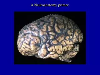
A Neuroanatomy primer. Gross surface anatomy of the human brain. References: Duvernoy, H. The Human Brain: Surface, Blood Supply, and Three-Dimensional Sectional Anatomy, 3 rd Edition, 1999: Absolutely the best atlas of the human brain and blood supply.

A Neuroanatomy primer.
E N D
Presentation Transcript
Gross surface anatomy of the human brain. References: Duvernoy, H. The Human Brain: Surface, Blood Supply, and Three-Dimensional Sectional Anatomy, 3rd Edition, 1999: Absolutely the best atlas of the human brain and blood supply. Nolte, J. The Human Brain 3rd Edition, Mosby Year Book, 1993: Good coronal slices and great in depth text on whole brain anatomy and motor pathways Damasio, H. Human Brain Anatomy in Computerized Images, Oxford University Press, 1995: Old but purely visual book that’s worth looking through A myriad of web sites – surf to your heart’s content! http://www.neuropat.dote.hu/anastru/anastru.htm - this site has great coronal images http://www.neuropat.dote.hu/atlas.html - same as the above site but with fantastic pathology pictures for those interested http://www.med.harvard.edu/AANLIB/home.html - nice neuropathology and movies of angiograms http://www.neuroguide.com/neuroimg_1.html#human_neuroanatomy – couldn’t get this one to work at time of writing this – but it looks interesting!
central (rolandic) sulcus frontal lobe parietal lobe occipital lobe temporal lobe sylvyan (lateral) sulcus Defining the lobes
Brodman’s areas Cytoarchitectonically defined brain regions – i.e., areas with the same physiological characteristics are grouped under a given number.
Development of Sulci Sulci appear at predictable points in fetal development with the most prominent sulci (e.g., Sylvian fissure) appearing first. Source: Ono, 1990
Comparative Neuroanatomy The complexity of sulci increased throughout evolution Source: Comparative Mammalian Brain Collection http://brainmuseum.org/
Major Sulci • Main sulci are formed early in development • Fissures are really deep sulci • Typically continuous sulci • Interhemispheric fissure • Sylvian fissure • Parieto-occipital fissure • Collateral sulcus • Central sulcus • Calcarine Sulcus • Typically discontinuous sulci • Superior frontal sulcus • Inferior frontal sulcus • Postcentral sulcus • Intraparietal sulcus • Superior temporal sulcus • Inferior temporal sulcus • Cingulate sulcus • Precentral sulcus • Other minor sulci are much less reliable Source: Ono, 1990
Interhemispheric Fissure - divides brain into 2 hemispheres
Sylvian Fissure (or lateral sulcus) -deep, mostly horizontal -insula (purple) is buried within it -separates temporal lobe from parietal and frontal lobes Sylvian Fissure
Parieto-occipital Fissure and Calcarine Sulcus Cuneus (pink) -visual areas on medial side above calcarine (lower visual field) Parieto-occipital fissure (red) -very deep -often Y-shaped from sagittal view, X-shaped in horizontal and coronal views Lingual gyrus (yellow) -visual areas on medial side below calcarine and above collateral sulcus (upper visual field) Calcarine sulcus (blue) -contains V1
Collateral Sulcus -divides lingual (yellow) and parahippocampal (green) gyri from fusiform gyrus (pink)
Cingulate Sulcus -divides cingulate gyrus (turquoise) from precuneus (purple) and paracentral lobule (gold)
ascending band of the cingulate Central, Postcentral and Precentral Sulci Central Sulcus (red) -usually freestanding (no intersections) -just anterior to ascending cingulate Precentral Sulcus (green) -often in two parts (superior and inferior) -intersects with superior frontal sulcus (T-junction) -marks anterior end of precentral gyrus (motor strip, yellow) Postcentral Sulcus (blue) -often in two parts (superior and inferior) -often intersects with intraparietal sulcus -marks posterior end of postcentral gyrus (somatosensory strip, purple)
Intraparietal Sulcus -anterior end usually intersects with inferior postcentral (some texts call inferior postcentral the ascending intraparietal sulcus) -posterior end usually forms a T-junction with the transverse occipital sulcus (just posterior to the parieto-occipital fissure - POF) -IPS divides the superior parietal lobule from the inferior parietal lobule (angular gyrus, gold, and supramarginal gyrus, lime) POF
Slice Views inverted omega = hand area of motor cortex
Superior and Inferior Temporal Sulci Superior Temporal Sulcus (red) -divides superior temporal gyrus (peach) from middle temporal gyrus (lime) Inferior Temporal Sulcus (blue) -not usually very continuous -divides middle temporal gyrus from inferior temporal gyrus (lavender)
Superior and Inferior Frontal Sulci Superior Frontal Sulcus (red) -divides superior frontal gyrus (mocha) from middle frontal gyrus (pink) Inferior Frontal Sulcus (blue) -divides middle frontal gyrus from inferior frontal gyrus (gold) orbital gyrus (green) and frontal pole (gray) also shown Frontal Eye fields lie at this junction
Medial Frontal -superior frontal gyrus continues on medial side -frontal pole (gray) and orbital gyrus (green) also shown
Arterial Blood Supply •Internal carotids supply hemispheres: •middle, anterior cerebral arteries, ophthalmic artery •vertebrals supply hemispheres, brainstem, spinal cord, cerebellum via numerous vessels. http://pathology.mc.duke.edu/neuropath/nawr/blood-supply.html#arteries great animation of blood supply
Circle of Willis •Internal carotid and vertebrals anastomoze in the Circle of Willis
Blood supply – lateral surface Middle cerebral artery – red Anterior cerebral artery – green Posterior cerebral artery – blue Veins - black frontoparietal parietal frontopolar superficial middle
Blood supply – medial surface • Anterior cerebral artery – green • Posterior cerebral artery – blue • Veins - black
Blood supply – inferior surface • Anterior cerebral artery – green • Posterior cerebral artery – blue • Veins - black
Aneurysms Angiogram - Aneurysm of ICA Blood vessels dissected - ACA aneurysm Aneurysm displaces hemisphere
Cerebral Vessel Infarcts Infarct of MCA Watershed infarct - fragile area at boundary of 2 vessels
Large draining veins. • Cerebral veins drain into veineous sinuses and into internal jugular • Superficial veins lie on surface of cortex and drain into superior sagittal sinus • Deep veins drain internal structures and empty into the straight sinus • Large draining veins can lead to artefacts in fMRI See Nolte, J. The Human Brain