Download
1 / 15
150 likes | 235 Vues
Understand female abdominal wall layers, reproductive organs, postpartum state, and menstrual cycle physiology. Explore anatomy of anterior abdominal wall and follow small-group discussions on relevant organs.

E N D
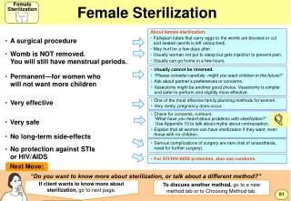
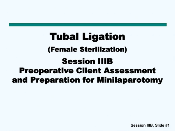
Tubal Ligation (Female Sterilization) Session IIIC: Basic Anatomy and Physiology of Female Reproductive System (including Anatomy of the Anterior Abdominal Wall)
SessionObjectives By the end of this session, the participant will be able to: • Identify the structures of the female abdominal wall • Identify the female reproductive organs relevant to minilaparotomy • Describe these organs in their postpartum state • Describe the basic physiology of the menstrual cycle
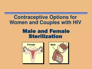
The Female Reproductive System Fallopian tube Ovary Clitoris Pubic hair Inner lip Opening for urine Outer lip Womb lining (endometrium) Vaginal opening Womb (uterus) Anus Other internal viscera of importance in surgery are the intestines, bladder, omentum, and ligaments such as the round, ovarian, and broad ligaments. Cervix Vagina
Small-Group Work • Discuss the location and functions of the following organs, including their relevance to minilaparotomy. Once you have completed the discussions, write your points on flipchart paper. • Time for group work: 10 minutes Group 1 • Vagina • Uterus • Fallopian tubes • Intestines Group 2 • Ovaries • Ligaments • Bladder • Omentum
Uterus: Positions a) Position of an anteverted uterus b) Positions of a retroverted and a retroflexed uterus Anteverted Uterus c) Insertion of elevator into the anteverted uterus Source: Engenderhealth 2003
Anatomical Changes During Pregnancy Uterus • The uterine wall undergoes physiological hypertrophy. • Blood vessels become enlarged. • All changes regress after delivery—known as involution. • Immediately postpartum, the uterine fundus is at the level of the umbilicus. Fallopian Tubes • The fallopian tubes are enlarged, more supple, engorged, edematous and friable
Postpartum Uterus Umbilicus Postpartum uterus Source Engenderhealth 2003
Involution of the Uterus Source Engenderhealth 2003
Postpartum Uterus: Incision Site a) Position of the uterus in relation to the Incision site in the subumbilicalminilaparotomy procedure b) Position of the incision site in the suprapubic minilaparotomy procedure Incision site Source: Engenderhealth 2014
Basic Female Reproductive Physiology—The Menstrual Cycle 2. Thickening of the womb lining(usually about 14 days long after ovulation) 1.Ovulation (usually occurs between days 7 and 21 of the cycle, often around day 14) X Egg Usually around 28 days long, but can range from 23 to 35 days. 4. Proliferative phase (This is first half of the menstrual cycle before ovulation when the lining of the uterus regenerates when the egg is also maturing) 3. Menstrual bleeding (period)(usually ranges from 2 to 7 days, often about 5 days)