Download
1 / 62
650 likes | 1.88k Vues
Small bowel obstruction &post operative ileus. Definition: mechanical or functional obstruction of the intestines, preventing the normal transit of the products of digestion. It is a medical emergency . Although many cases are not treated surgically, it is a surgical problem. Frequency:

E N D
Definition: mechanical or functional obstruction of the intestines, preventing the normal transit of the products of digestion. It is a medical emergency. Although many cases are not treated surgically, it is a surgical problem.
Frequency: Approximately 20% of patients admitted to the hospital with an acute abdomen have an intestinal obstruction (most common surgical disorder of small bowel)).
partial or complete, simple (ie, nonstrangulated) or strangulated. Strangulated obstructions (40%) are surgical emergencies which needsproper Dx and Rx.. If not diagnosed and properly treated, vascular compromise leads to bowel ischemia and further morbidity and mortality. Simple obstruction occludes the lumen only.(usually at one point). Strangulated obstruction impairs the blood supply and also leads to necrosis of the intestinal wall. Closed loop obstruction the lumen is occluded in at least 2 places(eg, in volvolus), is commonly ass. With strangulation
Mortality/Morbidity Mortality and morbidity are dependent on the etiology, the early recognition and correct diagnosis of obstruction. If untreated, strangulated obstructions cause death in 100% of patients. If surgery is performed within 36 hours, the mortality rate decreases to 8%. The mortality rate is 25% if the surgery is postponed beyond 36 hours in these patients.
Aetiology: can be classified into 3 main groups 'extraluminal' extrinsic (eg, adhesions, hernias, volvulus) intramural lesions in the bowel wall (eg, Crohn disease , tuberculosis, primary and secondary neoplasia, potassium strictures, radiation strictures, complications of surgical anastomosis) Intraluminal (eg, foreign bodies, bezoars, food bolus)
most common cause Adhesions (60%) related to previous surgery (within 4 weeks or decades later) or peritonitis. Adhesive bands occur between loops of bowel and the operative site causing acute angulation and kinking, The incidence parallels increasing number laparotomies developing countries. The second most common is an incarcerated hernia. A loop may enter any form of hernia and become obstructed narrow neck of a hernia, which compromises the caliber of the bowel . 1-external hernia (femoral, indirect inguinal, umbilical, incisional, epigastric, spigelian hernia) 2-internal hernia is clinically indistinguishable from obstruction resulting from postoperative adhesions.
Neoplasms 20 % ( intrinsic 3% extrinsic 17% ) Intrinsic neoplasms can either progressively occlude the lumen(small-bowel lymphoma and adenocarcinoma Lipomas, leiomyomas, and carcinoid tumors ) or ,more commonly, serve as leading point in intussusception (Any polypoid mucosal or submucosal lesion ). Extrinsic neoplasms: Secondary tumors ( gastric and colonic carcinomas, ovarian cancers, and malignant melanomas) may occasionally compromise the lumen of the small-bowel.
inflammatory bowel disease (5%) often causes obstruction when the lumen is narrowed by inflammation or fibrosis of the wall. volvulus (3%) results from malrotation of bowel loop around its mesenteric beds typically produces a closed loop of bowel with a pinched base, leading to intestinal obstruction with strangulation Small-bowel tuberculosis is not uncommon in certain parts of the world miscellaneous causes (2%). Intussusception: invagination of one loop of intestine to another is rarely encountered in adults (need leading point polyp or other intrluminal lesion. (colickly pain, blood per rectum, palpaple mass (intussuscepted segment).
Swallowed Forign bodies Bezoars A food bolus may occur, with indigestible vegetable material impacted in the terminal ileum. Patients with a food bolus will usually have undergone gastric outlet surgery. Gallstones may occur with a cholecystenteric fistula. Strictures may occur following ulceration induced by potassium tablets, nonsteroidal anti-inflammatory agents, and therapeutic irradiation for bladder or cervical cancer. An intramural hematoma may occur in cases of trauma or spontaneously in patients receiving higher doses of anticoagulant agents than are necessary.
Pathophysiology: Obstruction of the small bowel leads to proximal dilatation of the intestine due to accumulation of GI secretions and swallowed air. Swallowed air major source of gaseous distension (early) nitrogen is not well absorbed by the mucosa. Bacterial fermentation (later )other gases are produced partial pressure of nitrogen in the lumen are lowered; gradient of diffusion of nitrogen from blood to lumen. Large quantities of fluid from the extracellular space are lost into the gut ; and from the serosa into the peritoneal cavity. fluid fills the the lumen proximal to the obstruction; net secretion is enhanced mediators substances (endotoxin, prostaglandins) released from the luminal baceria are responsible. Reflexely induced vomiting accentuates the fluid and electrolytes deficit. Hypovolemia leads to multi-organ system failure and is the cause of death with non-strangulating obstruction.
In strangulated obstruction (eg, incarcerated hernia, volvolus) complete obstruction of the intestinal lumen as well as occlusion of the vascular supply( early venous drainage, then arterial supply).gangrenous bowel develops and might bleeds into the the lumen and into the peritoneal cavity and eventually it perforates. The luminal content of strangulated intestine (toxic mixture of bacteria,bacterial products,necrotic tissue and blood) Some of this fluid enter the circulation by way of lymphatics orby absorption from the peritoneal cavity, septic shock is the result. Note: Bacterial translocation from lumen to mesenteric L.N. and the bloodstream even in simple obstruction.
In general, the higher the level of obstruction, the less the distention and the more rapid the onset of vomiting. Conversely, in patients with a distal small-bowel obstruction, central abdominal distention may be marked and vomiting (feaculent) is, usually, a late feature (because the bowel takes time to fill). Colicky pain is most marked in patients with a distal obstruction. Hypotension and tachycardia suggest fluid depletion tenderness and leukocytosis suggest strangulation. In the early stages, bowel sounds are usually high-pitched, and they occur in frequent runs as the bowel contracts in an attempt to overcome the obstruction. A silent, tender abdomen suggests perforation or peritonitis, and it is a late sign
History partial or complete VS simple or strangulated. • Abdominal pain (characteristic with most patients) • Pain, often described as crampy and intermittent, is more prevalent in simple obstruction. • Often, the presentation the approximate location and nature of the obstruction. Usually, pain that occurs for a shorter duration of time and is colicky and accompanied by bilious vomiting may be more proximal. Pain lasting as many as several days, which is progressive in nature and with abdominal distention, may be typical of a more distal obstruction. • Changes in the character of the pain may indicate the development of a more serious complication (ie, constant pain of strangulated or ischemic bowel). • Nausea • Vomiting, which is associated more with proximal obstructions • In distal obstruction, (vomiting late,feaculent) • Diarrhea (an early finding) • Constipation (a late finding) as evidenced by the absence of flatus or bowel movements • Fever and tachycardia - Occur late and may be associated with strangulation • Virgin abdomen Previous abdominal or pelvic surgery, previous radiation therapy, or both (may be part of patient's medical history) • History of malignancy (particularly ovarian and colonic)
Examination: Vital signs: normal (early) Tachycardia, hypotension (late) Temperature: normal (simple) elevated (strangulation) Abdominal Ex: distension (more in distal). Mild tenderness Visible peristalsis Bowel sounds: hyperactive (early) hypoactive (late) Silent (peritonitis) Ex of hernias (incarcerated)
In strangulation: shock fever Cramping abd pain become severe continuos pain Abd. Tenderness and rigidity Silent abd Incarcerated hernia, abd. Mass (intussusceptum) Gross or occult blood Leukocytosis. acidosis note: no historical , physical or lab works entirely excludes the possibility of strangulation in complete SBO.
investigation: Essential laboratory tests Serum chemistries: Results are usually normal or mildly elevated. BUN level: If the BUN level is increased, this may indicate decreased volume state (eg, dehydration). Creatinine level: Creatinine level elevations may indicate dehydration. CBC: WBC count may be elevated with a left shift in simple or strangulated obstructions. Increased hematocrit is an indicator of volume state (ie, dehydration). Lactate dehydrogenase tests Blood gases analysis Urinalysis Type and crossmatch: The patient may require surgical intervention.
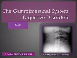
Imaging studies: Plain radiography: Obtain plain radiographs first for patients in whom SBO is suspected. At least 2, supine or flat and upright, are required Ladder-like pattern Dilated small-bowel loops with air fluid levels (>6) Absent or minimal colonic gas Intramural gas secondary to ischemia. This is a poor prognostic sign. Gallstone ileus presence of a calcified intraluminal stone (often in the terminal ileum) radiologic signs of a small-bowel obstruction above the Ileus, a gas in the biliary tree as a result of the cholecystoduodenal fistula.
Conventional barium follow-through examination and enteroclysis is valuable in detecting the presence of obstruction and in differentiating partial from complete blockages. useful when plain radiographic findings are normal in the presence of clinical signs of SBO or if plain radiographic findings are nonspecific. A delay in transit time on a conventional follow-through examination of greater than 12 hours is suggestive of an organic obstruction. Barium enema study ileocecal intussusception or other causes of ileocecal obstruction useful if a distal colonic obstruction cannot be excluded by using plain abdominal radiograph findings In children with intussusception, barium enema studies are not only diagnostic but possibly therapeutic as well.
dilated loops, stretching of the mucosal folds a narrowed segment ending in a beak (arrow )
. Multiple strictures and polypoid filling defectsvproximal small bowel deposits of non-Hodgkin lymphoma
ileocecal intussusception (carcinoid tumor of the terminal ileum)
double-contrast barium enema multiple fluid levels in the centrally placed S.B.
Stricture,shouldering of the terminal ileum caused by adenocarcinoma
Computed Tomography CT scans clearly demonstrate abnormalities of the bowel wall, the mesentery, the mesenteric vessels, and the peritoneum. useful in making an early diagnosis of strangulated obstruction particularly when clinical and radiographic findings are inconclusive. proved useful etiologies of SBO extrinsic causes such as adhesions and hernia from intrinsic causes such as neoplasms or Crohn disease. It also differentiates the above from intraluminal causes such as bezoars. about 90% sensitive and specific in detecting SBO. is the study of choice if the patient has fever, tachycardia, localized abdominal pain, and/or leukocytosis. It is capable of revealing abscess, inflammatory process, extraluminal pathology resulting in obstruction, and mesenteric Ischemia. enables the clinician to distinguish between ileus and mechanical small bowel in postoperative patients.
does not require oral contrast for the diagnosis of SBO because the retained intraluminal fluid serves as a natural contrast agent. • Obstruction is present if the small-bowel loop is greater than 2.5 cm in diameter dilated proximal to a distinct transition zone of collapsed bowel less than 1 cm in diameter. • A smooth beak indicates simple obstruction without vascular compromise; a serrated beak may indicate strangulation. • Bowel wall thickening indicates early strangulation. • Portal venous gas indicates early strangulation. • Pneumatosis indicates early strangulation. • useful in identifying abscesses, hernias, and tumors. • may be less useful in the evaluation of small bowel ischemia associated with obstruction
extrinsic mass compressing a loop of small bowel (desmoid t.)
Ultrasonography • Ultrasonography is less costly and less invasive than CT scanning. • It may reliably exclude SBO in as many as 89% of patients.
Management: • Continued NG suction: This provides symptomatic relief, decreases the need for intraoperative decompression, and benefits all patients. No clinical advantage to using a long tube (nasointestinal) instead of a short tube (NG) is observed. • Nonoperative treatment: A nonoperative trial of as many as 3 days is warranted for partial or simple obstruction. Provide adequate fluid resuscitation and NG suctioning Monitor urine output (foley cath) . Resolution of obstruction occurs in virtually all patients with these lesions within 72 hours. • administration of analgesia and antiemetic • Antibiotics are used to cover gram-negative and anaerobic organisms.
Surgical treatment: A strangulated obstruction is a surgical emergency. In patients with a complete SBO, the risk of strangulation is high and early surgical intervention is warranted. Patients with simple complete obstructions in whom nonoperative trials fail also need surgical treatment but experience no apparent disadvantage to delayed surgery. • Adhesions: Decreasing intraoperative trauma to the peritoneal surfaces can prevent adhesion formation. • Malignant tumor: Obstruction by tumor is usually caused by metastasis. Initial treatment should be nonoperative; surgical resection is recommended when feasible. • Inflammatory bowel disease: To reduce the inflammatory process, treatment generally is nonoperative in combination with high-dose steroids. Consider parenteral treatment for prolonged periods of bowel rest. Undertake surgical treatment, bowel resection, and/or stricturoplasty if nonoperative treatment fails.
Intra-abdominal abscess: CT-guided drainage is usually sufficient to relieve obstruction. • Radiation enteritis: If obstruction follows radiation therapy acutely, nonoperative treatment accompanied by steroids is usually sufficient. If obstruction is a chronic sequela of radiation therapy, surgical treatment is indicated. • Acute postoperative obstruction: This is difficult to diagnose because symptoms often are attributed to incisional pain and postoperative ileus. Treatment should be nonoperative. • Incarcerated hernia: Initially use manual reduction and observation. Advise elective hernia repair as soon as possible after reduction.
Indications for surgery • Absolute • Generalised peritonitis • Localised peritonitis • Visceral perforation • Irreducible hernia • Relative • Palpable mass lesion • 'Virgin' abdomen • Failure to improve • Trial of conservatism • Incomplete obstruction • Previous surgery • Advanced malignancy • Diagnostic doubt - possible ileus
Complications • Sepsis • Intra-abdominal abscess • Wound dehiscence • Aspiration • Short-bowel syndrome (as a result of multiple surgeries) • Death (secondary to delayed treatment)
Prognosis: With proper diagnosis and treatment of the obstruction, prognosis is good. Complete obstructions treated successfully nonoperatively have a higher incidence of recurrence than those treated surgically. Mortality and morbidity are dependent on the etiology, the early recognition and correct diagnosis of obstruction. If untreated, strangulated obstructions cause death in 100% of patients. If surgery is performed within 36 hours, the mortality rate decreases to 8%. The mortality rate is 25% if the surgery is postponed beyond 36 hours in these patients.
Paralytic ileus Background After abdominal surgery, a normal physiological ileus occurs. spontaneously resolves within 2-3 days the terms postoperative adynamic ileus or paralytic ileus are defined as ileus of the gut persisting for more than 3 days following surgery. Ileus occurs from hypomotility of the gastrointestinal tract in the absence of a mechanical bowel obstruction. This suggests that the muscle of the bowel wall is transiently impaired and fails to transport intestinal contents. This lack of coordinated propulsive action leads to the accumulation of both gas and fluids within the bowel.
the postoperative state is the most common scenario for ileus development. Frequently, ileus occurs after intraperitoneal operations, but it may also occur after retroperitoneal and extra-abdominal surgery. The longest duration of ileus is noted to occur after colonic surgery. The stomach regains activity in 1-2 days, and the colon regains activity in 3-5 days.and the small bowe within 24-48 hours
Causes of adynamic ileus • Sepsis • Drugs (eg, opioids, antacids, coumarin, amitriptyline, chlorpromazine) • Metabolic (eg, low potassium, magnesium, or sodium levels; anemia; hyposmolality) • Myocardial infarction • Pneumonia • Trauma (eg, fractured ribs, fractured spine) • Biliary and renal colic • Head injury and neurosurgical procedures • Intra-abdominal inflammation and peritonitis • Retroperitoneal hematomas
Clinical History • Patients with ileus typically present with vague, mild abdominal pain and bloating. • nausea, vomiting, and poor appetite. • Abdominal cramping is usually not present. • Patients may or may not continue to pass flatus and stool. • Hx previous operation
Physical • distended and tympanic abdomens, depending on the degree of abdominal and bowel distension. • may be tender. • A distinguishing feature is absent or hypoactive bowel sounds unlike the high-pitched sound of obstruction. • The silent abdomen of ileus reveals no discernible peristalsis or succussion splash.