CLINICOPATHOLOGICAL CONFERENCE PEDIATRICS
220 likes | 632 Vues
CLINICOPATHOLOGICAL CONFERENCE PEDIATRICS. Durante, Esperon, Espino, Fernando, Figuracion, Flores, Fong, Francisco, Francisco, Garcia, Garcia, Garcia, Garcia, Garcia, Garimbao . SUBJECTIVE. 10-year-old intermittent headache of 1 year duration vague frontal headaches

CLINICOPATHOLOGICAL CONFERENCE PEDIATRICS
E N D
Presentation Transcript
CLINICOPATHOLOGICAL CONFERENCE PEDIATRICS Durante, Esperon, Espino, Fernando, Figuracion, Flores, Fong, Francisco, Francisco, Garcia, Garcia, Garcia, Garcia, Garcia, Garimbao
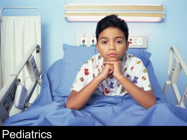
SUBJECTIVE • 10-year-old • intermittent headache of 1 year duration • vague frontal headaches • occur twice a week, usually in the late afternoons • diagnosed to have Iron Deficiency Anemia • prescribed with oral Iron preparation
SUBJECTIVE • projectile vomiting • non-villous, non-bloody • amounting to half a cup • occurs 2-3 times a day • did not experience tinnitus, gait disturbance, gastrointestinal, and urinary problems
SUBJECTIVE • allergic to shrimp • diagnosed with asthma last 2007 • family history of diabetes mellitus and hypertension
OBJECTIVE • slightly pale conjunctivae • + horizontal nystagmus • GCS 15 (E4V5M6) • positive for Romberg’s sign • no motor or sensory deficit • negative for Babinski sign, ankle clonus, nuchal rigidity, Kernig’s sign, and Brudzinski sign
COURSE IN THE WARDS • Admission • given Omeprazole 40 mg IV OD • to prevent irritation of the esophageal mucosa due to multiple bouts of vomiting • Ist HOSPITAL DAY • given Dexamethasone 2.5mg q6h • for the treatment of vasogenic edema associated with brain tumors • given Mannitol at 100 cc q6h • to decrease intracranial volume
COURSE IN THE WARDS • CSF analysis from ventricular drainage • 5 cc of clear, colorless fluid • pH of 7.5 • specific gravity of 1.010 • RBC 514 x 106 • WBC 1 x 10⁶, 100% lymphocytes • glucose of 4.7 mmol/L • protein 0.11 g/L • (-) Pandy’s
COURSE IN THE WARDS • 4TH HOSPITAL DAY • the patient underwent an operation • Ceftriaxone 750 mg IV was started and other medications were continued • 6th HOSPITAL DAY • Limited lateral eye movements on the left
COURSE IN THE WARDS • 7TH HOSPITAL DAY • Omeprazole IV and Dexamethasone IV were shifted to oral preparation • no episodes of vomiting were noted • MRI of the whole spine and liver function test • to evaluate for possible metastasis
PRIMARY IMPRESSION:MEDULLOBLASTOMA • Primarily considered due to: • Results of the patient’s CT scan (hyperdense lesion in the cerebellar vermis) • most common malignant hyperdense brain tumor arising in the cerebellar vermis • The patient’s age (10 y/o) • usually seen in 0-14 years of age
PRIMARY IMPRESSION:MEDULLOBLASTOMA • Presenting signs and symptoms • vague headache • vomiting • (+) Romberg sign • cranial nerve deficits
PRIMARY IMPRESSION:MEDULLOBLASTOMA • Incidence • accounts for 90% of embryonal tumors • 2% of all primary brain tumors • 18% of all pediatric brain tumors • predominately in males • majority occur in the midline cerebellar vermis
PRIMARY IMPRESSION:MEDULLOBLASTOMA • Signs and Symptoms • signs and symptoms of increased intracranial pressure and; • headache, nausea, vomiting, mental status changes, and hypertension • cerebellar dysfunction • ataxia, poor balance, dysmetria
PRIMARY IMPRESSION:MEDULLOBLASTOMA • Etiology and Pathogenesis • occur in the posterior fossa • 30–40% = chromosome 17p deletions • 10–20% = genetic loses on chromosomes 1q and 10p • 10% = abnormalities of chromosome 9p • arises from cerebellar stem cells • perivascular pseudorosette and Homer-Wright rosette formation
PLAN:Diagnostic Procedures • Laboratory studies • CBC, lectrolytes and liver and renal function tests • Imaging studies • CT scan, MRI, and bone scan • Other procedures • audiography or brainstem auditory-evoked response, • lumbar Puncture • bone marrow aspirate • biopsy and histologic study of the specimen
PLAN:Treatment • Surgery • to relieve cerebrospinal fluid buildup • to confirm the diagnosis by obtaining a tissue sample • to remove as much tumor as possible • Glucocorticoid treatment • to decrease the volume of edema surrounding brain tumors
PLAN:Treatment • ventriculostomy • to divert excess cerebrospinal fluid from the brain • radiation therapy • to reduce the number of left-over cells