Download
1 / 15
150 likes | 310 Vues
Regulation of lck degradation and refractory state in CD8 + cytotoxic T lymphocytes. Michael Uhlin, Maria G. Masucci, and Victor Levitsky * PNAS | June 28, 2005 | vol. 102 | no. 26 | 9264-9269. Date : 10/30/07 Name : Park Jong Chan. Abstract.

E N D
Regulation of lck degradation and refractory state in CD8+ cytotoxic T lymphocytes Michael Uhlin, Maria G. Masucci, and Victor Levitsky* PNAS | June 28, 2005 | vol. 102 | no. 26 | 9264-9269 Date : 10/30/07 Name : Park Jong Chan
Abstract • CD8+ cytotoxic T lymphocytes (CTLs)enter a refractory state termed activation- • induced nonresponsiveness (AINR) • 2. T cell receptortriggering results in rapid degradation of the lck through a • mechanism that is proteasome- and lysosome-independent,sensitive to cysteine • protease inhibitors. • 3. Pharmacologic blockade of lck degradation increased responsiveness of CTLs to • repeated antigenicchallenge. • 4. The development or maintenance of AINR was not affectedby exogenously • added IL-2, whereas IL-15 or IFN-a restored bothlck expression and • responsiveness of pre-activated CTLs. • 5. Lck degradation plays an important rolein the development of AINR in human • CTLs.
Introduction • CD4+ T cells : driven into an anergic state by T cell receptor (TCR) triggering in the absence of costimulation. • CD8+ CTLs : become unable to proliferate or produce IL-2 in response to specific stimulation subsequent to the primary TCR triggering combined with the engagement of co-stimulatory molecules. • 3. In mouse CTLs, AINR may be reverted by exogenous IL-2 • Human CTLs : it is not known whether the lymphokine acts in a similar • way on human CTLs • 5. Lck is an src-family protein kinase and One of the first molecules to be activated downstream of theTCR. • Acts asa tyrosine kinase and adaptor molecule • 6. A characteristic of Lck • lymphocytesinfiltrating tumors or circulating in the blood of patientswith chronic infections or inflammation
Materials and Method - Cell lines, CTL cultures and Clones EA/BK/ CAR: TCR structure of the IVT-peptide-specific • IVT : AVFDRKSVAK • AVT : IVTDFSVIK from EBV (Epstein–Barr virus) JAC-B2 : processes and presents the two peptides(IVT and AVT) endogenously. - Analysis of protein expression by immunoblotting - Inhibition of the proteasome and other protease. Epoxomycin(300nM), MG132(50-100uM), Leupeptin(100uM) Lactacystin(10uM), Z-LL-H(5-20uM) - FACS analysis - Plasmid preparation and Transfection of CTLs pcDNA3.1 , pcDNA3.1-lck - Analysis of T Cell Activation and Death.
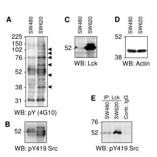
1. TCR Triggering Induces Lck Down-Regulation in Activated HumanT Cells Peptide treatment (1*10-7 M) Stimulation with APC Fig. 1. TCR triggering induces lck down-regulation in activated CD8+ T cells. • C1RA11 cells were pulsed with the AVF or IVT peptide (1 107 M), irradiated, and used to stimulate the indicated polyclonal CTL cultures. • (B) Expression of lck in CAR CTLs stimulated with the JAC-B2 LCL or L5 LCL. The latter carries mutations in the AVF and IVT • pitopes that prevent their resentation at the cell surface. • (C) T cell blasts were generated from peripheral blood mononuclear cells of five healthy blood donors by PHA activation. • On day 7 of culturing in IL-2 medium, 95% of cells in every culture were CD3CD8 as determined by FACS analyses • Down-regulationof lck in response to TCR triggering is not clon- or donor- • dependent and can be induced by physiological amounts of specific antigen.
2. Specific Activation of CTLs Induces Rapid and Persistent Down-Regulationof Lck Expression. 56kDa 59kDa Fig. 2. Kinetics of lck down-regulation in IVT-specific CTL • First lane band : Lck polypeptide of 56kDa expressed in resting CTLs • lane second bend : phosphorylated form of lck • Phosphorylated form of lck was not dectable from 8h postactivation.
3. Degradation of Lck Is Blocked by Cysteine Protease Inhibitors. Fig. 3. AID of lck is proteasome- and lysosome-independent but sensitive to inhibition by MG132 • Immunoblotting of lysates of BK289 cells with ubiquitin-, lck-, p53-, or actin-specific Abs. • Effect of epoxomycin on the steady-state levels of lck and p53. • (C) Effects of epoxomycin on AID of lck or p53 were analyzed and expressed • Down-regulation of lck was not affected by the presence of epoxomycin or lactacystyin during T cell triggering. • Inhibitor cause accumlation of ubiquitinated proteins and p53 • Steady-state levels of lck were decreased in the presence of proteasome inhibitor.
Fig. 3. AID of lck is proteasome- and lysosome-independent but sensitive to inhibition by MG132 (D) Effect of MG132 on lck degradation assessed at the indicated concentrations. (E) Quantification of the effect of MG132 at a concentration of 100 µM (F) Control or activated CTLs were cultured in the presence or absence of NH4Cl. Immunoblotting of total cell lysates with lck-, IL-15Ra -, or actin-specific Abs. • MG132 blocked down-regulation of lck in a does-dependent manner. • The NH4Cl induced accumulation of the IL-15 receptor a-cahin in nonactivated CTLs and further increased its up-regulation after CTL activation. • NH4Cl did not affect AID of lck, indicating that lysomal enzymes are not involved in this process.
Fig. 4. The calpain inhibitor Z-LL-H blocks AID of lck. BK bulk CTLs were activated by IVT-pulsed APCs for 4 h in the absence (control) or presence of Z-LL-H at the indicated concentrations. • Effect Z-LL-H on the AID of lck, ZAP-70, or zeta chain tested by immunoblotting with the relevant Abs • Intensities of lck, ZAP-70, or zeta chain-specific bands expressed as percentage expression relative to band intensities in samples of unstimulated CTLs. • The AID of lck in CTLs pre-incubated with and activated in the presence of L-ZZ-H was strongly inhibited. • The steady-state levels of z-chain and ZAP-70 increased
Is Lck Degradation necessary for Efficient T Cell Activation??? [ One possible interpretation ] lck degradation is required for efficient TCR signal transduction and T cell activation. The effect of Z-LL-H on the capacity of BK CTLs to kill C1RA11 cells and produce IL-2 or IFN-r in response to stimulation with IVT pulsed APCs. Result : None of these parameters of T cell activation was negatively affected by Z-LL-H. A comparable decrease of cell recovery was observed in BK bulk CTL cultures activated by IVT-pulsed APCs in the presence or absence of Z-LL-H, whereas the release of IFN-r was slightly increassed in the presence of the inhibitor. Lck Degradation Is Not a Prerequisite for Efficient T Cell Activation.
5. Down-Regulation of Lck Through Degradation Plays a Role in the Development of AINR in CTLs. Wild type : IVT Partial agonistic variants : Y5 : F to Y substitution A8 : I to A substitution Wild type : 34% to 25% decrease Fig. 5. Responsiveness of CTLs correlates with lck expression, and treatment with Z-LL-H prevents the development of AINR in specific CTLs. • BK bulk CTLs were stimulated during the indicated periods of time with C1RA11 cells preincubated with the indicated synthetic peptides. The expression of lck was accessed by immunoblotting and quantified as described above. • (B) CTLs preactivated by using the indicated peptides were re-stimulated with IVT pulsed APCs after 48 h, and their proliferation was evaluated by a [3H]thymidine incorporation assay. * A strong correlation was observed between the level of lck down-regulation induced by a given peptide and the extent to which responsiveness of the CTLs was inhibited.
(C and D) BK bulk CTLs were activated for 2 h on plastic plates with absorbed CD3-specific Ab, transferred to a clean plate, and cultured for 48 h either in the absence or presence of 20uM Z-LL-H. Lck expression in these cells was evaluated by immunoblotting. (D) Cells were then activated by IVT-peptide pulsed APCs, and their proliferation was evaluated by a [3H]thymidine incorporation assay. * The presence of L-ZZ-H during the first CTL triggering inhibited lck degradation as well as the development of AINR in these cells.
Purpose : To directly access the role of lck down-regulation in AINR Fig. 6. Transfection of pcDNA3.1-lck into refractory CTLs enhances their capacity to produce IFN-r in response to specific stimulation * First triggering -> Transfection with pcDNA3.1-lck or control pcDNA3.1 plasmid-> Overnight culture -> Re-stimulation -> measuring the IFN-r
6. IFN- and IL-15 Reconstitute Lck Expression and Abrogate the Development of AINR in Specific CTLs. • Fig. 7. IL-15 and IFN-a increase lck expression after specific activation and restore responsiveness in preactivated CTLs. • The BK bulk CTLs were activated with peptide-pulsed APCs. • Capacity of CTLs to proliferate in response to the secondary challenge as evaluated by a [3H]thymidine incorporation assay. • Physiological signals can reconstitute lck expression after TCR triggering. • IVT-specific CTLs cultured in the presence of exogenously added IL-15 or IFN- did not develop AINR in response to TCR triggering and proliferated after the secondary challenge almost as efficiently as control cells
Discussion • we provide evidence implicating activation-induced degradation of lck in the development of AINR in CTLs. • modulation of lck expression is one of the key molecular events responsible for the well documented capacity of IL-15 and INF- to promote both primary and memory CTL responses. • low levels of lck correlate with AINR in CTLs and that reconstitution of lck expression and reversion of AINR can be achieved by pharmacologic agents and lymphokines paves the way to the development of new approaches aiming to improve the immunologic control of tumors and infections.