Download
1 / 24
310 likes | 1.05k Vues
Life of a Cell. The Cell Cycle and Mitosis. Mitosis (copy this information on the back of your Mitosis cut & paste page). Process that divides the nucleus of a cell Cell division that results in two identical diploid “daughter” cells.

E N D
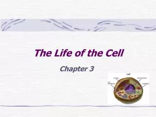
Life of a Cell The Cell Cycle and Mitosis
Mitosis(copy this information on the back of your Mitosis cut & paste page) • Process that divides the nucleus of a cell • Cell division that results in two identical diploid “daughter” cells. • Each daughter cell receives the exact same # and type of chromosomes • Mitosis occurs for the purpose of: • Reproduction (unicellular organisms only) • Growth and repair/maintenance of tissues(multicellular organisms) • Consists of 4 phases: • Prophase, Metaphase, Anaphase, and Telophase (PMAT) • Followed directly by cytokinesis (divison of cytoplasm)
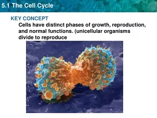
The Cell Cycle • Consists of: • Interphase • Mitosis/ Cytokinesis Mitosis is a small part of the cycle; a cell spends almost all of it’s time in interphase. Most cells don’t continually go through the cell cycle; many stay in interphase, without preparing to divide, for long periods (G0).
Instructions for taking notes • For each phase (interphase, prophase, etc.) copy the information from the slide onto the cut & paste page. • If only certain phrases are bolded/underlined, copy those. • If noting is bolded/underlined, copy it ALL. • Study the pictures provided of the phases. Label certain structures on your cut & paste page (such as centrioles, chromosomes, nucleus, etc.) • You don’t have to label these things in every picture. But, be sure some pictures have labels.
Interphase(write the description below on your cut & paste page, under the first and last pictures) • NOT a “resting” phase for the cell. The cell carries out normal metabolic processes (like respiration, protein synthesis, etc.) and prepares for cell division (if given signals that cell division is needed). • Stages of interphase include: • G1: Cell growth • S: DNA is replicated (copied) • G2: growth and preparations for cell division
cell wall Interphase (Plant) Notice that the nucleus is clearly visible (with a nucleolus), but no chromosomes are visible. The DNA is in the invisible form of chromatin.
Interphase (Animal) Cell membrane Nuclear membrane
Centrosomes (w/centrioles, if it’s an animal cell) Watch the animation. Pause it when it begins to talk about metaphase. Animation: http://highered.mcgraw-hill.com/sites/0072495855/student_view0/chapter2/animation__mitosis_and_cytokinesis.html Prophase: Mitosis Begins(Copy the info below beneath the 2 pictures of prophase) • Chromatin coils into chromosomes (becomes visible) • Chromosomes consist of 2 chromatids • Centrosomes migrate to opposite sides of cell; spindle fibers form and attach to chromosomes • Nuclear membrane dissolves (disappears) You have 2 pictures of prophase: an “early” prophase and a “late” prophase. The picture below is of “early” prophase. As prophase proceeds the centrosomes would end up on opposite poles and the chromosomes would be distinct.
Prophase (Plant) Chromatin is condensing into chromosomes. The nuclear membrane is dissolving.
Prophase (Animal) Chromosomes
Centromere Centrosome/ Centriole Metaphase Illustration *Chromosomes consist of two chromatids
Metaphase: Organizing the Chromosomes(Copy the info below beneath the picture of metaphase) • During metaphase, chromosomes are pulled by spindle fibers to the mid-line (“equator”) of the cell. • They align “single file”. • Organizing the chromosomes before separation will ensure that each new cell will receive a full set of chromosomes (genetic info).
Metaphase (Plant) Chromosomes lined up along the “equator” of the cell.
Centriole Spindle Fibers Chromatid (now is also called a chromosome) Centromere Watch the animation. Pause it when it begins to talk about telophase. Animation: http://highered.mcgraw-hill.com/sites/0072495855/student_view0/chapter2/animation__mitosis_and_cytokinesis.html Anaphase Illustration(Copy the info below beneath the 2 pictures of anaphase) • In Anaphase: • Chromatids separate at the centromere, becoming individual chromosomes • Chromosome now = 1 chromatid • Spindle fibers shorten, pulling chromatids to opposite poles of cell
Anaphase (Plant) Chromatids
Anaphase (Animal) Chromatids Centriole
Telophase Illustration(Copy this under the picture of telophase.) • In telophase: • Nuclear membrane forms again; 2 nuclei are formed. • Chromosomes uncoil into long strands of chromatin • Cytokinesis begins
Telophase (Plant) Nuclear membrane forming Cytokinesis beginning to seperate the 2 nuclei into 2 separate cells. Chromatin
Telophase (Animal) Cytokinesis beginning to seperate the 2 nuclei into 2 separate cells. Nucleus
Cleavage furrow Watch the remainder of the animation. Animation: http://highered.mcgraw-hill.com/sites/0072495855/student_view0/chapter2/animation__mitosis_and_cytokinesis.html Cytokinesis Illustration(Copy this under the picture of telophase.) • In cytokinesis: • The cytoplasm divides between the daughter cells • Happens by cleavage furrow (animal) or formation of cell plate (plant) • Produces 2 daughter cells
Cleavage Furrow Cytokinesis (Animal)
Quiz yourself! • Go to: http://school.discoveryeducation.com/quizzes6/muskopf/mitosis1.html • On the back of your cut & paste page, write your answers to the quiz questions, in complete sentences.