Download

1 / 72
751 likes | 1.85k Vues
Gestational Trophoblastic Disease (GTD). Department of Obs. & Gyn., First Hospital of Xi ’ an Jiaotong University Gao Shang Feng. Introduction. What is GTD ? It is a rare kind of disease in which abnormal trophoblastic proliferation occurs.

E N D
Gestational Trophoblastic Disease (GTD) Department of Obs.& Gyn., First Hospital of Xi’an Jiaotong University Gao Shang Feng
Introduction What is GTD ? • It is a rare kind of disease in which abnormal trophoblastic proliferation occurs. • It is too among the rare human malignancies that can be cured even in the presence of widespread metastases.
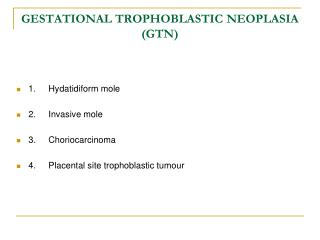
Which does it include? • It includes a spectrum of interrelated tumors, including • hydatidiform mole (HM) • invasive mole (IM) • Choriocarcinoma (CH) • Placental-site trophoblastic tumor (PSTT, borderline, very rare)
Relationship of HM. IM. CH hydatidiform therapeutic or molespontaneous abortion term pregnancy ectopic invasion mole choriocarcinoma.
What is GTT (Gestational trophoblastic tumor)? GTT is all GTD except hydatidiform mole. They has its unique pathologic characteristics and biological behavior. Even the most malignant case can be cured by chemotherapy.
Hydatidiform mole It is a neoplastic proliferation of the trophoblast in which the terminal villi are transformed into vesicles filled with clear viscid material.
It is usually benign but has malignant potentiality. • Incidence: • south east Asia is 1/500-600 • the US and Europe:1/500-2000 • China:1/1238
Classification It is divided into two classification • complete hydatidiform mole • partial hydatidiform mole
complete hydatidiform mole(CHM): • the entire uterus filled with abnormal vesicles, no signs of fetus.
partial hydatidiform mole • partial hydatidiform mole with evidence of a conceptus.
Etiology Though it is not known a number of associated factors have been noted: • the absence of fetal circulation; • dietary protein deficiency • viral infection; • age:>45 years women are 10 times more likely to develop HM than those younger
abnormal fertilization process: • the fertilization of a normal ovum with a duplicated haploid sperm:46XX • the fertilization of an empty egg by two sperms(dispermy):46XY
Chromosomes complete hydatidiform moles • Cytogenetic studies have demonstrated that complete hydatidiform moles usually have a 46xx karyotype, and the molar chromosomes are entirely of paternal origin. • Complete moles appear to arise from an ovum that has been fertilized by a haploid sperm, which then duplicates its own chromosomes, and the ovum nucleus may be either absent or inactivated
Although most complete moles have a 46xx chromosomal pattern, approximately 10% have a 46xy karyotype. • Chromosomes in a 46xy complete mole also appear to be entirely of paternal origin, but in this circumstance, an apparently empty egg is fertilized by two sperm.
. partial hydatidiform mole • partial moles usually have a triploid karyotype (69 chromosomes ), with the extra haploid set of chromosomes derived from the father. • When a fetus is present in conjunction with a partial mole, it usually exhibits the stigmata of triploidy, including growth retardation and multiple congenital malformations.
complete hydatidiform mole pathology Complete moles lack identifiable embryonic or fetal tissues, and the chorionic villi exhibit generalized hydatidiform swelling and diffuse trophoblastic hyperplasia.
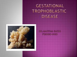
Gross we see a mass of vesicles, vary in size, grape-like with stems, blood and clot filling the inter-vesicle space
partial hydatidiform mole It are characterized by the following pathologic features : • Chorionic villi if varying size with focal hydatidiform swelling and cavitation. • It contain identifiable embryonic or fetal tissues.
Gross we see a mass of vesicles, vary in size, grape-like and identifiable embryonic or fetal tissues.
Microscopic ‘ • trophoblastic proliferation. • hydropic degeneration of the stroma. • absence of blood vessels or extreme scantiness of blood vessels.
Normal trophoblastic partial hydatidiform mole complete hydatidiform mole
trophoblastic proliferation is considered the most important single criteria. • Ovaries respond to hCG stimulation ,30-50% theca-lutein cysts develop, bilateral
Clinical course It has eight of symptoms and physical signs.
amenorrhea because it is a pregnancy. • vaginal bleeding after a period of amenorrhea (average 12 weeks) may continue intermittently for several weeks---profuse bleeding---anemia and infection. • abdominal cramps
abnormally enlarged and soft uterus in about half the cases, the uterus growth is rapid, it is larger than the dates suggest.
ovarian cyst torsion when we do pelvic examination adnexal masses may be found. it is theca lutein cyst in about one third of the cases
severe and early –onset PIH (Pregnancy Induced Hypertension syndrome) • hyperthyroidism plasma thyroxin concentration elevates • exaggerated early pregnancy symptoms nausea, vomit etc
Diagnosis suspicion: • abnormal bleeding after amenorrhea • inappropriately enlarged uterus; • absence of fetal heart sounds or could not feel fetal parts by palpation between 16-20th week • hyperemesis gravidarum • bilateral ovarian cysts
serum hCG monitor an unusually high titer of chorionic gonadotropin, especially after the one-hundredth day of pregnancy, help to confirm the diagnosis of HM.
Ultrasonography: It is a reliable and sensitive technique for the diagnosis of complete molar pregnancy. Because the chorionic villi exhibit diffuse hydatidiform swelling. Complete moles produce a characteristic vesicular sonographic pattern, usually referred to as a “snowstorm” pattern.
Ultrasonography may also contribute to the diagnosis of partial molar pregnancy by demonstrating focal cystic spaces in the placental tissues and an increase in the transverse diameter of the gestational sac.
Differential diagnosis • abortion; • multiple pregnancy; • polyhydramnios
Treatment • the uterus should be evacuated as soon as possible after the diagnosis is made.(by suction curettage) • suction; • oxytocin administration:we can use blood transfusion or/and fluid infusion.it is used to decrease the size of the uterus;
tissue sent for histology: it should be routine practice with all cases of incomplete miscarriage; • acute pulmonary complications
total abdominal hysterectomy in older multiparas hysterectomy may be indicated.
management of theca-lutein cysts these tumors should not be excised because they regress after the trophoblastic tissue has been removed.
chemotherapy HM don’t need usually chemotherapy because HM is benign disease.
㈧Follow-up examinations • follow up mode in the 2 years after discharge • on each follow-up check, the following should be addressed
symptom abnormal vaginal bleeding, cough, hemoptysis signs of metastasis • pelvic examination • hCG evaluation • B-ultrasound • chest X-ray film
contraceptive method required for 1-2 years • condom is recommended. • IUD (intrauterine device)and pills are contraindicated for their potentiality of causing abnormal vaginal bleeding.
Ask question • What is the etiology of GTD? • What is the classification of HM? • What is the main pathologic changes of HM? • What is the clinical course of HM? • How Follow-up examinations is we?
About 80% of the cases of HM have a benign course. one-half of patients become pregnant subsequently. about 16% of HM become invasion moles and some 2.5% progress intochoriocarcinoma
Introduction • Invasion Mole arises from HM • it has malignant potentialities, invades the myometrium and penetrates the uterine wall, extends into the broad ligament or peritoneal cavity.
in half or more of all cases invasive mole metastasizes through the peripheral circulation to distant sites, mostly to the lung.
Pathologic findings • excessive trophoblastic proliferation and invasiveness • the degree of anaplasia is variable: completely benign---highly malignant
differentiation between invasive mole and choriocarcinoma lies in whether the villous pattern is preserved: • if we see villi, it must be invasion mole; • if we can’t see villi, it is choriocarcinoma.