Download
1 / 21
220 likes | 695 Vues
NOVAK 34. Gestational Trophoblastic Disease. 부산백병원 산부인과 R1 손영실. # Hydatidiform Mole (H-mole). INDEX. 1. Epidemiology 2. Complete Versus Partial Hydatidiform Mole 3. Clinical Features 4. Natural History 5. Diagnosis 6. Treatment 7. Follow-up. EPIDEMIOLOGY. ◎ Risk Factors

E N D
NOVAK 34. Gestational Trophoblastic Disease 부산백병원 산부인과 R1 손영실
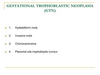
INDEX 1. Epidemiology 2. Complete Versus Partial Hydatidiform Mole 3. Clinical Features 4. Natural History 5. Diagnosis 6. Treatment 7. Follow-up
EPIDEMIOLOGY ◎ Risk Factors - low nutritional and socioeconomic factors - low dietary intake of carotene - vitamin A deficiency - maternal age older than 35 years - use of oral contraceptive - history of irregular menstruation
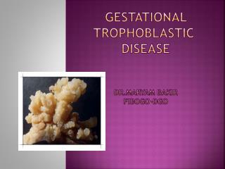
COMPLETE VERSUS PARTIAL HYDATIDIFORM MOLE (on the basis of gross morphology, histopathology, and karyotype)
COMPLETE VERSUS PARTIAL HYDATIDIFORM MOLE 1. Complete H-mole ◎ Pathology - lack embryonic or fetal tissues - chorionic villi → generalized hydatidiform swelling & diffuse trophoblastic hyperplasia ◎ Chromosomes - usually have a 46,XX - molar chromosomes are entirely of paternal origin - ovum nucleus may be either absent or inactivated - 10% : 46,XY
Empty ovum 23X 23X 46XX Endoreduplication Homozygous 23X Empty ovum 46XX 23X Heterozygous 23X Empty ovum 46XY 23Y 46YY Non-viable gamete COMPLETE VERSUS PARTIAL HYDATIDIFORM MOLE
COMPLETE VERSUS PARTIAL HYDATIDIFORM MOLE 2. Partial H-mole ◎ Pathology ① Chorionic villi of varying size with focal hydatidiform swelling, cavitation, and trophoblastic hyperplasia ② Marked villous scalloping ③ Prominent stromal trophoblastic inclusions ④ Identifiable embryonic or fetal tissues ◎ Chromosome - generally have a triploid karyotype (69 chromosomes) - extra haploid set of vhromosome usually is derived from the father - 90 ~ 93% : triploid
23X 23X 69XXX 23X 23Y 23X 69XXX 23Y 23X 23X 69XXX 23Y 69YYY Non-viable gamete COMPLETE VERSUS PARTIAL HYDATIDIFORM MOLE
CLINICAL FEATURES 1. Complete H-mole ① Vaginal bleeding - most common symptom - 97% → 84% - molar tissue separate from decidua & disrupt maternal vessels → large volumes of retained blood may distend endometrial cavity ② Excessive uterine size - relative to gestational age - one of classic signs of complete mole - expanded by both chorionic tissue & retained blood - generally associated with elevated levels of hCG
CLINICAL FEATURES ③ Preeclampsia - observed in 27% of patients with complete mole - associated with HBP, proteinuria, and hyperreflexia - eclamptic convulsion rarely occur - preeclampsia develops almost in patients with excessive uterine size & markedly elevated hCG ④ Hyperemesis gravidarum - occurred in 25% of women with complete mole - particularly with excessive uterine size & markedly elevated hCG
CLINICAL FEATURES ⑤ Hyperthyroidism - 7% of women in complete mole - Sx : tachycardia, warm skin, and tremor - Dx : serum free T4, T3 - If suspected before surgery, β-adrenergic blocking agent should be administered (to prevent many of the metabolic and cardiovascular complication of thyroid storm)
CLINICAL FEATURES ⑥ Trophoblastic embolization - 2% of women in complete mole - Sx : chest pain, dyspnea, tachypnea, tachycardia & severe respiratory distress (during and after molar evacuation) ⑦ Theca lutein ovarian cysts - 50% of patients with complete mole - result from high hCG levels, cause ovarian hyperstimulation - after molar evacuation, cysts normally regress spontaneously within 2 to 4 months
CLINICAL FEATURES 2. Partial H-mole • Do not have the dramatic clinical feature • In general, patients have the sign and symptoms of incomplete or missed abortion • partial mole can be diagnosed after histologic review of the tissue obtained by curettage
NATURAL HISTORY 1. Complete H-mole - have a potential for local invasion(15%) and metastasis(4%) (after molar evacuation) - following signs ① hCG level > 100,000 mIU/ml ② excessive uterine enlargement ③ theca lutein cysts 6cm in diameter - patients with any one of these signs → high risk 2. Partial H-mole - 4% of patients : persistent tumor, usually nonmetastatic, chemotherapy is required to achieve remission
DIAGNOSIS - Ultrasonography is a reliable and sensitive technique for diagnosis - Characteristic vesicular ultrasonographic pattern : snowstorm pattern (honey-comb appearance)
TREATMENT 1. Hysterectomy - if the patient desires surgical sterilization, hysterectomy may be performed - the ovaries may be preserved, even though prominent theca lutein cysts are present - hysterectomy does not prevent metastasis, so, still required f/u hCG levels
TREATMENT 2. Suction Curettage - preferred method of evacuation, for patients who desire to preserve fertility - the following steps ① oxytocin infusion : before induction of anesthesia ② cervical dilatation : retained blood in endometrial cavity may be expelled during dilatation ③ suction curettage : uterus may decrease dramatically in size ④ sharp curettage : performed to remove any residual molar tissue
TREATMENT 3. Prophylactic Chemotherapy prevented metastasis reduced the incidence and morbidity of local uterine invasion - single course of actinomycin D at time of evacuation - useful in the management of high-risk complete mole
FOLLOW - UP 1. Human Chorionic Gonadotropin (hCG) - monitored with weekly of hCG levels until these levels are normal for 3 consecutive weeks - followed by monthly until levels are normal for 6 consecutive months 2. Contraception - patients should be used effective contraception during the entire interval of hCG f/u - oral contraceptive may be used safely