Download
1 / 25
270 likes | 562 Vues
Ectopic pregnancy, spontaneous abortion and gestational trophoblastic disease. SUFIA HUSAIN Pathology KSu , Riyadh April 2014. Objectives. At the end of this lecture, the student should be able to:

E N D
Ectopic pregnancy, spontaneous abortion and gestational trophoblastic disease. SUFIA HUSAIN Pathology KSu, Riyadh April 2014
Objectives At the end of this lecture, the student should be able to: Understand the pathology and predisposing factors of ectopic pregnancy and spontaneous abortion. Know the clinical presentation and pathology of hydatidiform mole and choriocarcinoma.
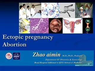
Ectopic Pregnancy • Definition: Ectopic pregnancy is defined as implantation of a fertilized ovum in any site other than the endometrium of the uterine cavity. As many as 1% of pregnancies are ectopic. • Sites: • Over 90% of ectopic pregnancies occur in the fallopian tubes (tubal pregnancy). • Other sites of ectopic pregnancy include the ovaries, abdominal cavity and uterine cervix.
Ectopic Pregnancy Clinical features • A woman with an ectopic tubal pregnancy may present with pelvic pain or abnormal bleeding following a period of amenorrhoea. • The majority will present as an emergency with tubal rupture and hemorrhagic shock. Diagnosis • Clinical: abdominal/pelvic ultrasound shows mass (gestational sac) within fallopian tube, plus positive hCG levels • Microscopic: placental tissue or fetal parts
Risk factors for ectopic pregnancy Tubal ectopic pregnancy is the more common type and any factor that retards passage of the ovum through the tubes predisposes to tubal ectopic pregnancy. In about half of the cases, it is due to chronic inflammation and scarring in the oviduct. The risk factors are as follows: • Pelvic inflammatory disease/infections/salpingitis is one of the most common cause. Organisms like Neisseriae gonorrhea and chlamydia infect the reproductive organs. Pelvic infection may alter tubal function, damaged ciliary activity , cause tubal obstruction and pelvic adhesions with scarring and distortion of the fallopian tubes. Women who have had pelvic infections have a five times greater risk of ectopic pregnancy. • Abdominal/pelvic surgery or tubal ligation surgery. • Intrauterine tumors and endometriosis may also hamper passage of the ovum. • Smoking may contribute to decreased tubal motility by damaging ciliated cells or it may predisposing them to pelvic inflammatory disease (due to the impaired immunity in smokers). • Congenital anomaly of the tubes. • In-utero diethylstilbestrol (DES) exposure increases the risk of ectopic pregnancy due to abnormal tubal morphology.
Risk factors for ectopic pregnancy • History of previous ectopic pregnancy • History of multiple sexual partners is associated with an increased risk of ectopic pregnancy because of the increase chance of pelvic inflammatory disease . • Intrauterine device users are at higher risk of having an ectopic pregnancy if pregnancy occurs. • History of infertility: there is higher risk of ectopic pregnancy in the infertile population (whether treated or not). This may be due to the underlying infertility related issues or fertility drugs and treatments, such as in vitro fertilization. In vitro fertilization has been associated with an increased risk of ectopic pregnancy including cervical pregnancies • NOTE: In the other 50% of tubal pregnancies, no anatomic cause is evident. Ovarian pregnancies probably result from rare instances in which the ovum is fertilized just as the follicle ruptures. Gestation within the abdominal cavity occurs when the fertilized egg drops out of the fimbriated end of the oviduct and implants on the peritoneum.
Spontaneous abortion (SAB)/ Miscarriage • Miscarriage is the spontaneous end of a pregnancy at a stage where the embryo or fetus is incapable of surviving. • Miscarriages that occur before the sixth week of gestation are medically termed early pregnancy loss or chemical pregnancy. • Miscarriages that occur after the sixth week LMP are medically termed clinical spontaneous abortion. • 10-25% of all clinically recognized pregnancies will end in miscarriage. • Most miscarriages occur during the first 13 weeks of pregnancy.
Causes of SAB/Miscarriage: The cause of a miscarriage cannot always be determined. Miscarriages can occur for many reasons. Some of these causes include genetic, uterine or hormonal abnormalities like diabetes, collagen vascular disease (e.g. SLE), reproductive tract infections, and congenital (present at birth) abnormalities of the uterus Most miscarriages occur during the first trimester. Chromosomal abnormalities of the fetus are the most common cause of early miscarriages. The causes are as follows 1. Chromosomal abnormalities: Half of the 1st trimester miscarriages have abnormal chromosomes. This number drops to 20% with 2nd trimester miscarriages. Chromosomal abnormalities also become more common with aging, and women over age 35 have a higher rate of miscarriage than younger women. A pregnancy with a genetic problem has a 95% probability of ending in miscarriage.
Causes of SAB/miscarriage 2. Hormonal problems: there is an increased risk of miscarriage with Cushing’s Syndrome, thyroid disease and polycystic ovary syndrome (PCOS). Diabetes generally can be well managed during pregnancy. Good control of blood sugars during pregnancy is very important. However, if the diabetes is insufficiently controlled, there is increase risk of miscarriages and also the baby can have major birth defects. It also has been suggested that inadequate function of the corpus luteum in the ovary (which produces progesterone necessary for maintenance of the very early stages of pregnancy) leads to progeterone deficiency which may lead to miscarriage. It is termed as "luteal phase defect’’. 3. Infections:. infections by Listeria monocytogenes, Toxoplasma gondii, parvovirus B19, rubella, herpes simplex, cytomegalovirus and lymphocytic choriomeningitis virus etcare associated with an increased risk of pregnancy loss. 4. Maternal health problems e.g. Collagen vascular diseases are illnesses in which a person's own immune system attacks their own organs e.g. systemic lupus erythematosus and antiphospholipid antibody syndrome are associated with an increased risk of miscarriage are 5. Lifestyle (i.e. smoking, drug use, malnutrition, excessive caffeine and exposure to radiation or toxic substances)
Causes of SAB/miscarriage 6. Abnormal structural anatomy of the uterus can also cause miscarriages. In some women there can be a tissue bridge (uterine septum), that acts like a partial wall dividing the uterine cavity into sections. The septum is not well suited for placental attachment and growth. Therefore, an embryo implanting on the septum would be at increased risk of miscarriage. Structural abnormalities in the uterus like fibroids can uncommonly interfere with the embryo implantation and the embryo's blood supply, thereby causing miscarriage 7. Maternal age: SABs increase after age 35 due to ovum abnormalities 8. Maternal trauma 9. Others like invasive surgical procedures in the uterus during pregnancy e.g amniocentesis and chorionc villus sampling.
Causes of SAB/miscarriage Diagnosis: A miscarriage can be confirmed via ultrasound and by the examination of the passed tissue microscopically for the products of conception. The products of conception include chorionic villi, trophoblasts, fetal parts, and background gestational changes in the endometrium. Genetic tests may also be performed to look for chromosomal anomalies.
Gestational Trophoblastic Disease (GTD) • Gestational trophoblastic disease comprises a heterogeneous group of related lesions arising from abnormal proliferation of placental trophoblasts. • GTD are divided into • benign non-neoplastic lesions • hydatidiform moles • neoplastic lesions • Most GTD produces the beta subunit of human chorionic gonadotropin (HCG).(note: serum HCG is also elevated in normal and ectopic pregnancybut it is markedly increased is GTD. In addition the serum HCG levels in GTD continue to rise even after 14th weeks in contrast to normal preganacy in which the HCG levels drop after 14 weeks gestation. • Most women who have had gestational trophoblastic disease can have normal pregnancies later. • The maternal age over 40 years found to have a 5.2-fold increased risk of trophoblastic disease in compression to the mothers below the age of 35 years.
Types of GTD There are various histologically distinct subtypes of GTD: • Benign non-neoplastic trophoblastic lesions — These are diagnosed as an incidental finding on an endometrial curettage or hysterectomy specimen. They are: • Exaggerated placental site • Placental site nodule • Hydatidiform mole — result from abnormalities in fertilization. They are essentially benign, but carry an increased risk of developing malignant choriocarcinoma. They are: • Complete hydatidiform mole • Partial hydatidiform mole • Invasive mole/chorioadenomadestruens (some invasive moles that do not resolve spontaneously are considered neoplastic) • Gestational trophoblastic neoplasia (GTN) — is a group of tumors with the potential for local invasion and metastases. In contrast to other more common malignancies, GTN is curable in majority (85-100%) of cases. They are: • Choriocarcinoma • Placental site trophoblastic tumor • Epithelioid trophoblastic tumor
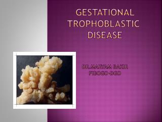
Hydatidiform Mole • It is the abnormal fertilization resulting in an abnormal placenta due to excess of paternal genes. It is caused by abnormal gametogenesis and fertilization. • It is the most common form of gestational trophoblastic disease; occurs in 1/1,000-2,000 pregnancies • In it there is enlarged and odematous placental villi filling the lumen of the uterus. • Passage of tissue fragments, which appear as small grapelike masses, is common. The serum hCG concentration is markedly elevated, and serial determinations disclose rapidly increasing levels. • Risk factors: • maternal age: girls younger than 15 years of age and women over 40 are at higher risk. • Ethnic background: incidence is higher in Asian women than among white women • Women with a prior hydatidiform mole have a 20-fold greater risk of a subsequent molar pregnancy than the general population. • There are 2 types of hydatidiform mole (HM). • Complete HM • Partial HM
Complete HM • Complete mole results from fertilization of an empty ovum that lacks maternal DNA. Most commonly, a haploid (23,X) set of paternal chromosomes duplicates to 46,XX. The characteristic feature is complete lack of maternal chromosomes. • It is a genetically abnormal placenta with hyperplastic trophoblasts, without fetus or embryo. • Symptoms: with increased abdominal swelling (rapid increase in uterine size) mistaken for normal pregnancy but the uterus is disproportionately large for stage of pregnancy. In addition patient has some vaginal bleeding, severe nausea and vomiting. There is elevated HCG levels. • Uterus is distended and filled with swollen/large villi. No embryo, or fetal tissue is present. Grossly it looks like a bunch of grapes. • Chromosomal analysis shows 46XX karyotype and all the chromosomes come from the male/paternal side i.e. it is an androgenetic pregnancy with no maternal DNA.
Complete HM. • The ultrasound will often show a “cluster of grapes” appearance or a “snowstorm” appearance, signifying an abnormal placenta. • Treatment : Evacuation of uterus by curettage and sometimes chemotherapy. With appropriate therapy cure rate is very high. • Complications: uterine hemorrhage, uterine perforation, trophoblastic embolism, and infection. Few patients develope an invasive mole. The most important complication is the development of choriocarcinoma, which occurs in about 2% of patients after the mole has been evacuated.
Partial Mole (PM) Partial hydatidiform moles have 69 chromosomes (triploidy gestation), of which one haploid set (23,X) is maternal and two haploid (23,X+23X=46X) sets are paternal in origin. This results from fertilization of a normal single ovum/egg (23,X) by two normal spermatozoa, each carrying 23 chromosomes, or by a single spermatozoon that has not undergone meiotic reduction and bears 46 chromosomes(the pregnancy has too much paternal DNA). It is a genetically abnormal placenta with a mixture of large and small villi with slight hyperplasia of the trophoblasts, filling the uterus. In contrast to a complete mole, embryo/fetal parts may be present. The fetus associated with a partial mole usually dies after 10 weeks' gestation, and the mole is aborted shortly thereafter. It almost never evolves into choriocarcinoma. Grossly the genetically abnormal placenta has a mixture of large chorionic villi are mixed with normal-appearing smaller villi.
Partial Mole (PM) It makes up 15–35% of all moles Uterine size usually small or appropriate for gestational age Serum hCG levels are elevated but not as high as complete mole. Chromosomal analysis of partial moles shows 69XXY (i.e. 3 haploid sets also called as triploidy). Treatment : Evacuation of uterus by curettage and sometimes chemotherapy. Prognosis: Risk for development of choriocarcinoma very low. Follow-up is mandatory.
2. Invasive Mole Invasive mole is when the villi of a hydatidiform mole extends/infiltrates into the myometrium of the uterus. The mole sometime tends to enter dilated veins in the myometrium, and a times through the vascular channels spread to distant sites, mostly the lungsbut the villi do not penetrate out of the blood vessels in which they are lodged, and therefore death from such spread is unusual. It occurs in about 15% of complete moles and rarely in partial mole. Can cause hemorrhage and uterine perforation.
3. Choriocarcinoma Definition: Malignant tumor derived from normal or abnormal placental tissue, composed of a proliferation of malignant cytotrophoblast and syncytiotrophoblast, without villi formation. It is an aggressive malignant neoplasm. It is characterized by highly increased serum concentration of HCG. Choriocarcinomas are aneuploidic. It spreads early via blood to the lungs and other organs. Responds well to chemotherapy About half the choriocarcinoma are preceded by complete hydatidiform mole . Others are preceded by partial mole (rare), abortion, ectopic pregnancy and occasionally normal term pregnancy.