Download

1 / 71
720 likes | 782 Vues
Learn about the classification, diagnosis, and treatment of gestational trophoblastic neoplasia including hydatidiform mole, choriocarcinoma, and placental site trophoblastic tumor.

E N D
Staging, Classification and Treatment of GestationalTrophoblastic Disease Assoc. Prof. Gazi YILDIRIM, M.D. Yeditepe University, Medical Faculty Dept of Ob&Gyn
Objectives • To define • Molpregnancy • Malignantgestationaltrophoblastictumors • Tolearn • Management of GTN
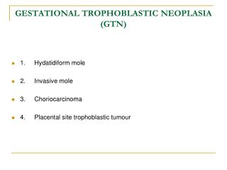
Classification • Histological classification • Partial mole • Complete mole • Invasive mole • Metastatic mole • Choriocarcinoma • Placental-site trophoblastlc tumour • Epithelioid trophoblastic tumour • Clinical classification of gestational trophoblastlc neoplasla • Non-metastatic • Metastatlc • Low risk • High risk
Types of GTD Benign • Hydatidiform mole/molar pregnancy (complete or incomplete) Malignant • Invasive mole • Choriocarcinoma (chorioepithelioma) • Placental site trophoblastic tumor
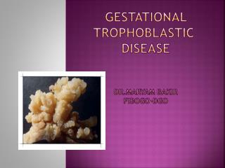
HYDATIDIFORM MOLE (MOLAR PREGNANCY) • Histologically chorionic villi consist of trophoblastic proliferation and Edema of villous stroma • Moles usually occupy the uterine cavity • The absence or presence of a fetus or embryonic elements has been used to describe them as completeand partial moles
COMPLETE HYDATIDIFORM MOLE • Chorionic villi transform into a mass of clear vesicles • Vesicles vary in size • The histological structure typically shows: 1. Hydropic degeneration and swelling of the villous stroma. 2. Absence of blood vessels in the swollen villi. 3. Proliferation of the trophoblastic epithelium to a varying degree 4. Absence of fetus and amnion.
Chromosomal composition in 85 percent of complete molar pregnancies is 46,XX, with both chromosomes being of paternal origin • This phenomenon is termed androgenesis. • Typically, the ovum has been fertilized by a haploid sperm, which then duplicates its own chromosomes after meiosis. • The chromosomes of the ovum are either absent or inactivated. • Occasionally, the chromosomal pattern in a complete mole may be 46,XY due to dispermic fertilization
COMPLETE MOLE A single sperm fertilizes an empty ovum, with duplication of the 23X haploid set of chromosomes, giving rise to a homozygous diploid complete mole. Empty ovum 46XX 23X Complete Mole (46XX diploid) Two sperms with two independent haploid sets of chromosomes fertilize an empty ovum, producing a dyspermic complete mole with either 46XX or 46XY karyotype. Empty ovum 46XX or 46XY 23X 23X or Y Complete Mole (46XX or 46XY, diploid) Modified from Cheung, 1995
PARTIAL HYDATIDIFORM MOLE • Hydatidiform changes are focal and less advanced • Some element of fetal tissue is seen • There is slowly progressive swelling within the stroma of characteristically avascular chorionic villi • Other vascular villi with a functioning fetal-placental circulation • Karyotype typically is triploid 69,XXX, 69,XXY, or 69,XYY • These are each composed of one maternal and two paternal haploid sets of chromosomes • Nontriploid partial moles have been reported • The nonviable fetus of a triploid partial mole typically has multiple malformations • Abnormal growth is common • 82 percent of fetuses had symmetrical growth restriction.
PARTIAL MOLE 23Y 23X 23X 23X 23X 69XXY 23Y Dyspermy 23X/23Y or 23X/23X Partial Mole (69XXY, or 69XXX, or 69XYY triploid) Fertilization of a normal 23X haploid ovum by two sperms, producing a triploid partial mole with either 69XXY, 69XXX or 69XYY karyotype Modified from Cheung, 1995
A twin gestation of a complete mole and a normal fetus and placenta is sometimes misdiagnosed as a diploid partial mole • Twin pregnancies consisting of a normal fetus and a complete mole have a substantively increased risk of developing gestational trophoblastic neoplasia • Generally, gestational trophoblastic neoplasia follows 20 percent of complete moles, whereas it develops in only 0.5 percent of women after a partial mole • The risk of choriocarcinoma arising from a partial mole is also low
FEATURES OF PARTIAL AND COMPLETE MOLE Complete mole Partial mole Fetal or embryonic tissue absent present Hydatiform swelling of chorionic villi extensive focal Trophoblastic hyperplasia extensive focal Scalloping of chorionic villi absent present Trophoblastic stromal inclusions absent present Karyotype 46XX (90%); Triploid (69 XXY) 46XY (10%) Cohn DE, Herzog TJ. Curr Opin Oncol 2000 Sep; 12(5):492-6
INCIDENCE AND RISK FACTORS • Hydatidiform mole develops in approximately 1 in 1000 pregnancies in the United States and Europe. • Although it has been reported to be more frequent in other countries, especially in parts of Asia • The role of gravidity, estrogen status, oral contraceptives, and dietary factors in the risk of gestational trophoblastic disease is unclear
Age. incidence of molar pregnancy highest in women aged 15 years or younger, and those aged 45years or older. • In the latter group, the relative frequency of the lesion is at least 10 times greater than that at ages 20 to 40 years
Previous Mole. Women with molar pregnancies are at increased risk of developing either a complete or a partial mole in a future pregnancy • Recurrent moles 1.3 percent • Oocyte problem leads to molar development
CLINICAL COURSE. • Bleeding. • Vary from spotting to profuse hemorrhage. • There may be considerable hemorrhage concealed within the uterus. • Iron-deficiency anemia is common
Uterine Size. • Growing uterus often enlarges more rapidly than usual • Exceeding in about half of cases that expected from the gestational age. • The uterus may be difficult to identify precisely by palpation because of its soft consistency. • Fetal Activity. • Typically no fetal heart motion is detected. • There may be incomplete molar degeneration in the placenta accompanied by a living fetus.
Gestational Hypertension. • Because hypertension caused by pregnancy is rarely seen before 24 weeks, preeclampsia that • develops before this gestational age may be from hydatidiform mole or extensive molar degeneration. • Hyperemesis. • Significant nausea and vomiting may develop. • Thyrotoxicosis. • Plasma thyroxine levels in women with molar pregnancy are often elevated, but clinically apparent hyperthyroidism is unusual • Hyperthyroidism is identified in about 2 percent. • Serum free thyroxine is elevated as the consequence of the thyrotropin-like effect of hCG
Embolization. • Variable amounts of trophoblastic cells escape from the uterus into the venous outflow at the time of molar evacuation • The volume may be such as to produce signs and symptoms of acute pulmonary embolism or edema. • This tissue can subsequently invade the pulmonary parenchyma to establish metastases.
DIAGNOSTIC FEATURES • To summarize, the clinical and diagnostic features of a complete hydatidiform mole are: 1. Continuous or intermittent brown or bloody discharge evident by about 12 weeks and usually not profuse. 2. Uterine enlargement out of proportion to the duration of pregnancy in about half of the cases. 3. Absence of fetal parts and fetal heart motion. 4. Characteristic ultrasonographic appearance. 5. Serum hCG level higher than expected for the stage of gestation. 6. Preeclampsia-eclampsia developing before 24 weeks. 7. Hyperemesis gravidarum.
PROGNOSIS • Mortality from molar pregnancies has been practically reduced to zero by prompt diagnosis and appropriate therapy. • Earlier evacuation has not reduced the 20 percent risk for gestational trophoblastic neoplasia
TREATMENT • Hydatidiform mole treatment consists of two phases. The first is immediate evacuation of the mole, The second is subsequent evaluation for persistent trophoblastic proliferation or malignant change. • Computed tomography or magnetic resonance imaging to evaluate the liver or brain is not performed routinely.
Prophylactic Chemotherapy. • Controversial • Toxicity from prophylactic chemotherapy may be significant • Chemoprophylaxis may be considered in women with high-risk complete moles, particularly if serum hCG testing is unavailable or follow-up is impossible
Vacuum Aspiration. • Treatment of choice for hydatidiform mole • Cervical dilating agents may be necessary • The cervix may be further dilated under anesthesia to a diameter sufficient to allow insertion of a suction curet. • After most of the molar tissue has been removed by aspiration, oxytocin is given. • Intraoperative ultrasonographic examination may help document that the uterine cavity has been emptied.
Oxytocin, Prostaglandins, and Hysterotomy. • Labor induction rarely is used for evacuation of hydatidiform moles. • Hysterectomy. • If no further pregnancies are desired, hysterectomy may be preferred to suction curettage. • Hysterectomy does not eliminate recurrent disease, it appreciably reduces likelihood
FOLLOW-UP EVALUATION OF MOLAR PREGNANCY 1. Prevent pregnancy for a minimum of 6 months using hormonal contraception. 2. Monitor serum hCG levels every 2 weeks. Serial measurement of serum hCG is important to detect trophoblastic neoplasia, 3. Chemotherapy is not indicated as long as these serum levels continue to regress. A rise or persistent plateau in the level demands evaluation for gestational trophoblastic neoplasia An increase signifies trophoblastic proliferation that is most likely malignant unless the woman is again pregnant. 4. Once the hCG level falls to a normal level, test the patient monthly for 6 months; then follow-up is discontinued and pregnancy allowed. • Estrogen-progestin contraceptives or depot-medroxyprogesterone usually are used to prevent a subsequent pregnancy during the period of surveillance.
GESTATIONAL TROPHOBLASTIC NEOPLASIA • Also called malignant gestational trophoblastic disease, • Refers to • invasive mole, • choriocarcinoma, • placental site trophoblastic tumor. • Any of these may follow molar pregnancy or normal pregnancy, or develop after abortive outcomes, including ectopic pregnancy.
ETIOLOGY • Gestational trophoblastic neoplasia almost always develops with or follows some form of pregnancy. • Approximately • 50% of cases follow a hydatidiform mole, • 25% follow an abortion, • 25% percent develop after an apparently normal pregnancy.
PATHOLOGY • The diagnosis of gestational trophoblastic neoplasia is made primarily by persistently elevated serum hCG levels.
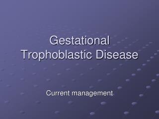
Definition A malignant form of GTD which can develop from a hydatidiform mole or from placental trophoblast cells associated with a healthy fetus ,an abortion or an ectopic pregnancy.
Definition • Characterized by abnormal trophoblastic hyperplasia and anaplasia , absence of chorionic villi
Microscopic image of choriocarcinoma absence of chorionic villi
Common Sites for Metastatic Gestational Trophoblastic Tumors
Definition This term is applied to a molar pregnancy in which molar villi grow into the myometrium or its blood vessels, and may extend into the broad ligament and metastasize to the lungs, the vagina or the vulva.
Invasive mole: the tissue invades into the myometrial layer. No obvious borderline, with obvious bleeding.
Definition • Placenta Site Trophoblastic Tumor is an extremely rare tumor that arised from the placental implantation site • Tumor cells infiltrate the myometrium and grow between smooth-muscle cells
Placental Site Trophoblastic Tumor. • Trophoblastic neoplasia arises from the placental implantation site following a normal term pregnancy, spontaneous or induced abortion, ectopic pregnancy, or molar pregnancy • Histologically predominantly cytotrophoblastic cells, and immunohistochemical staining reveals many prolactin-producing cells and few gonadotropin-producing ones • Thus, serum hCG levels may be normal to elevated. • Bleeding is the main presenting symptom. • hPL is high (human Placental Lactogen)