Pediatric Umbilical Abnormalities
Pediatric Umbilical Abnormalities. Scott Nguyen MD Mount Sinai School of Medicine Dept of Surgery. Abnormalities of Umbilical Cord. Umbilical abnormalities result from failure of umbilical ring to close or persistence of umbilical structures

Pediatric Umbilical Abnormalities
E N D
Presentation Transcript
Pediatric Umbilical Abnormalities Scott Nguyen MD Mount Sinai School of Medicine Dept of Surgery
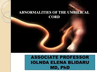
Abnormalities of Umbilical Cord • Umbilical abnormalities result from failure of umbilical ring to close or persistence of umbilical structures • Understanding embryology of cord is essential in understanding the pathophysiology of umbilical abnormalities
Embryology • 6th wk – midgut loop elongates and herniates out through umbilical cord • Midgut rotates 270 degrees • Returns to abdomen by 10th wk • Anterior abdominal wall progressively closes leaving only umbilical ring
Umbilical Abnormalities • Urachal Abnormalities • Vitelline Duct Abnormalities • Umbilical Hernia • Omphalitis • Delayed Cord Separation
Bladder forms from ventral portion of cloaca Bladder descends into pelvis w/ urachus connecting apex to umbilicus Usually urachus involutes to a fibrous cord – median umbilical ligament Urachal formation
Urachal abnormalities • failure of obliteration of urachus resulting complete or partial patency of urachus • < 1/1000 live births • inflammation or drainage from umbilicus • US, CT, contrast studies, or injection of dye into tract can confirm diagnosis
Patent Urachus (50%) Urachal cyst (30%) Urachal sinus (15%) Vesicourachal diverticulum (5%)
Studies • Catherization of tract and injection of dye • Voiding cystourethrogram • US
Usually assx until infected Rarely become infected in newborn period, usu manifests as young adult Urachal Cyst
Infected Urachal cyst • Fever, voiding symptoms, midline hypogastric tenderness, mass, UTI • May drain into bladder or umbilicus • Rarely can rupture into preperitoneal tissues or peritoneal cavity • Cultures - Staph Aureus
Infected Urachal cyst - treatment • Incision and drainage • Percutaneous drainage • Complete surgical excision of all urachal tissue • 30% recurrence if only drainage • Staged approach limits amount of bladder resected
Becomes symptomatic when infected Tx – drainage and resection of urachal tissue Urachal Sinus
Blind sac at bladder apex Mostly assx Urachal Diverticulum
Vitelline Duct • Vitelline Duct is connection between midgut and yolk sac • Usually involutes in 7th – 9th weeks
Meckel’s Diverticulum • contains ectopic gastric or pancreatic mucosa • In 2% of population • 2 feet from ileocecal valve, antimesenteric border • Majority of symptomatic < 2yrs old
Presentation • Painless GI Bleeding (50%) • Bowel Obstruction (30%) • Inflammation – diverticulitis (20%)
GI Bleeding • Most common cause of bleeding in children • Painless, massive, usually self resolving • Due to mucosal ulceration from acid secretion
Bowel Obstruction • Due to intussusception, diverticulum is the lead point • Sudden severe pain out of proportion to physical exam • Hydrostatic Barium enema diagnostic, rarely therapeutic
Meckel’s Diverticulitis • Sx like appendicitis • Result of lumenal obstruction, bacterial invasion, progressive inflammation • Ectopic gastric mucosa predisposes • 30% incidence of perforations • Higher risk of peritonitis
Treatment • Surgical Resection without removal of ileum • V shaped incision at base • resection of involved segment of ileum w/ primary anastamosis
Vitelline Umbilical fistula • Umbilical polyp • May drain enteric contents • Fistulogram shows communication w/ bowel
Umbilical hernia • Protrudes • Rarely incarcerates • Incidence 10-25% infants • 6-10x higher incidence in Black infants • More in girls, premature • Assoc w/ Down’s Synd, Beckwith-Wiedemann synd, hypothyroidism, mucopolysaccharidosis