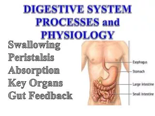

Digestive Physiology
E N D
Presentation Transcript
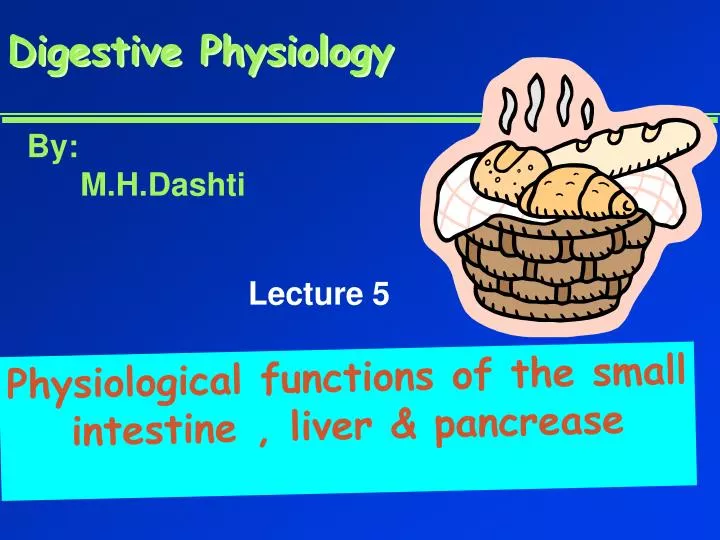
Digestive Physiology By: M.H.Dashti Lecture 5 Physiological functions of the small intestine , liver & pancrease
Anatomy of the Small Intestine • 20 feet long----1 inch in diameter • Large surface area for majority of absorption • 3 parts • duodenum---10 inches • jejunum---8 feet • ileum---12 feet • ends at ileocecal valve
Secretions of the small intestine many villi on surface of intestine : 2 types of glands 1- Brunner’s glands- compound mucus glands, Secreting Alkaline mucus -Stimulated by parasympathetic -Inhibited by sympathetic -?stress related ulceration 2- crypts/glands of Lieberkuhn between villi Exocrine from Enterocyte Endocrine: Secretin (S) , cck ( I ),GIP ( K ) ,VIP ( v ) ,Motilin ( mo ) Digestive enzymes not secreted from small intestine except enterokinase secreted from duodenal mucosa
Crypt of Lieberkuhn Villus under local nervous control some minor hormonal control (VIP ,secretin, CCK ) Mucous cells (simple goblet cells) Enterocyte (absorptive cells with brush border) Endocrine cells (secrete hormones for control of gut function) Paneth cell (have Secretory granules of lysozyme)
HCO3- Secretin Secretin Fat H+ S cells stomach Bile S.I.
CCK Cholecystokinin Stomach stomach duodenum I cells S.I. Bile + Peptides Amino acids , pancreas Fat Enzymes Insulin Glucagon
Other intestinal hormones • GIP (Gastric Inhibitory peptide ) from k cells • Stimulated by carbohydrates in duodenum • Stimulate insulin secretion • VIP ( Vasoactive Intestinal Peptide ) • Stimulated by H+ in duodenum • Inhibit gastric secretion & Stimulate intestinal secretion • VIPoma • Motilin • Stimulated by H+ in duodenum • Co-related to Migratory Myoelextric Complex ( MMC )
Small intestine motility • Weak peristalsis in comparison to the stomach---chyme remains for 3 to 5 hours • Segmentation---local mixing of chyme with intestinal juices---sloshing back & forth
Bile Production • 0.5-1 Lof bile/day is secreted • yellow-green in color & pH 7.6 to 8.6 • Components: water • Cholesterol , FA & Lecithin • bile salts = Na & K salts of bile acids • bile pigments (bilirubin) • Alkalinephosphatase • Gall bladder concentrates bile by absorbing water & Na-cl
Bile production & enterohepatic circulation Bilirubin is produced by the metabolism of Heme& is transported to the liver by albumin
Gallstones • Calcium - bilirobinate due to glucoronidase activity of bacteria • Cholesterol stones due to increased Cholesterol/PLratio
Pancreatic Secretions & their Regulation • Both endo. & exocrine Secretions are under neural & hormonal control • Regulated in 3 phases • Secretin • increased sodium bicarbonate release from ducts • CCK • increased digestive enzymes & hormones ( I & G ) release ( EGSF ) • GIP • Stimulated by fatty acids & sugar in S.I. • causes increased insulin release (EISF )
Delivers Inositol tri-phosphate Adenylate cyclase Cholera toxin Regulation of pancreatic acinar secretion
Sites of pancreatic secretions • Acinar fluid is isotonic & its ionic composition resembles the plasma • Spontaneous secretion of interalobular ducts have greater K+ & HCO3- than plasma • Secretin stimulated secretion from the cells of extra lobular ducts are still more rich in HCO3-
H2O H2O K+ Cellular mechanisms for HCO3- rich secretion by pancreatic acinar cells
H2O • Cellular mechanisms for HCO3- rich secretion by pancreatic extra lobular ductal cells • Stimulated by secretin , cck & Ach
Ionic concentration of pancreatic juice as a function of its flow rate