Download
1 / 32
320 likes | 500 Vues
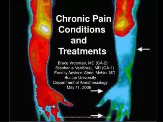
Chronic Pain Conditions and Treatments. Bruce Vrooman, MD (CA-2) Stephanie VanKraaij, MD (CA-1) Faculty Advisor: Abdel Mehio, MD Boston University Department of Anesthesiology May 11, 2006.

E N D
Chronic Pain Conditions and Treatments Bruce Vrooman, MD (CA-2) Stephanie VanKraaij, MD (CA-1) Faculty Advisor: Abdel Mehio, MD Boston University Department of Anesthesiology May 11, 2006 http://www.thermogramcenter.com/Images_files/RSD%20arms.jpg
Conditions: Myofascial Pain Intercostal Neuralgia Postherpetic Neuralgia CRPS Type I : RSD Type II: Causalgia Cancer Pain Procedures: Triggerpoint Injections Neurostimulation: TENS Spinal Cord Stimulators Peripheral Nerve Stimulators Neurolytic Blocks Sympathetic Nerve Blocks, TENS, Drug Treatments Physical Therapy Drug Treatments Regional Anesthesia Sympathetic Blockade Stellate, Lumbar, and Celiac Ganglion Blocks IV Regional Blockade Epidural Blockade Brachial Plexus Blockade Neurostimulation Drug Treatments Celiac Sympathetic Plexus Block Neurosurgery Outline Note: ASA Board Exam content outline is listed at the end of this presentation. Back Pain conditions and treatments will be covered by the lecture next week
Condition: Myofascial Pain • Signs and Symptoms: • Tenderness of triggerpoints in skeletal muscles • Appearance of ropy bands of skeletal muscle • Jump Sign: trigger area palpated and patient “jumps” from the pain • Scapulocostal syndrome: trigger point medially and superior to upper scapula. • May cause referred pain in occiput, shoulder, or chest. • Gluteal muscle involvement: pain to posterior thigh and calf, mimicking S1 radiculopathy Source: Stoelting and Miller
Treatment: Myofascial Pain • Physical Therapy • Diagnostic Injection • 0.5% lidocaine or 0.25% bupivacaine with cortisol 25 mg • Provides analgesia • Confirms diagnosis • Allows for initiation of PT • U/S, TENS, or vapocoolant spray as alternatives to provide analgesia. Source: Stoelting andMiller
Procedure: Trigger Point Injections Most frequent locations of myofascial trigger points Examples of the three directions in which trigger points (Xs) may refer pain (red). (A) Peripheral projection of pain from suboccipital and infraspinatus trigger points. (B) Mostly central projection of pain from biceps brachii trigger points with some pain in the region of the distal tendinous attachment of the muscle. (C) Local pain from a trigger point in the serratus posterior inferior muscle. Peripheral Mostly Central Local http://www.aafp.org/afp/20020215/653.html
Procedure: Trigger Point Injections Cross-sectional schematic drawing of flat palpation to localize and hold the trigger point (dark red spot) for injection. (A, B) Use of alternating pressure between two fingers to confirm the location of the palpable nodule of the trigger point. (C) Positioning of the trigger point halfway between the fingers to keep it from sliding to one side during the injection. Injection is away from fingers, which have pinned down the trigger point so that it cannot slide away from the needle. Dotted outline indicates additional probing to explore for additional adjacent trigger points. The fingers are pressing downward and apart to maintain pressure for hemostasis. http://www.aafp.org/afp/20020215/653.html
Neurostimulation Treatment Overview • TENS • Spinal Cord Stimulation • Not initial treatment • Considered after failure of oral medications for control of pain with peripheral neuropathic pain or pain arising from spinal cord • Peripheral Nerve Stimulation • For patients with peripheral mononeuropathy who have responded to diagnostic sequence of local neural blockade and stimulation trial Source: Stoelting and Miller
Procedure: TENS www.healingtools.tripod.com/ BS.jpg www.1-800-medical.com/.../ paintech/elechart.gif
TENS • Used for persistent pain from: • Back surgery • Peripheral nerve injury • Phantom limb pain • Occasionally postherpetic neuralgia • Patient-activated delivery of pulsed electrical current to skin over painful areas • Current activates large afferent fibers, causing stimulation of inhibitory dorsal horn neurons and release of endorphins and preventing spasm • Activates descending inhibitory system for preventing transmission of pain • Clinical application of “Gate Theory” Source: Stoelting and Miller
TENS • Gate Theory: • A-beta fibers inhibits transmission of pain impulses via A-delta and C fibers • Biochemical mechanisms involved • TENS increases CSF levels of Substance P and 5-hydroxytryptamine • Maximally-conformable paresthesia at site of pain to produce effective analgesia Source: Stoelting and Miller
Electricity triggers response blocking the transmission of pain signaling to the brain Used to treat chronic trunk and limb pain Electrodes are placed in the epidural space. Three components: Power source Electrode leads External controller Short noninvasive surgical procedure With power on, targeted nerves stimulated, changing pain messages: eg. Paresthesia rather than pain. Trial stiumulator used for one week to see if patient tolerates this Usually placed if conservative therapy (medications, ESI or nerve blocks) has failed www.lowbackpain.com/ spinalCordStim.shtml Procedure: Spinal Cord Stimulation bss.ewi.utwente.nl/ research/neurostimulation www.lowbackpain.com/ spinalCordStim.html
Condition: Intercostal Neuralgia • Definition • S/P thoracotomy or rib fracture • Usually several weeks, yet may persist for several years • Treatment • Neurolytic blocks with alcohol or phenol • Local anesthetic intercostal or paravertebral nerve blocks • PT to start in pain-free interval Source: Stoelting and Miller
Procedure: Neurolytic Blocks • Usually for palliative care, such as from terminal cancer, because of recovery of pain sensation in weeks. • Phenol • injected in epidural or subarachnoid spaces. • may cause denervation hypersensitivity pain if by peripheral nerve • 5% to 20% phenol for peripheral nerves • Painless at injection, delayed onset of neurolysis, hyperbaric • Alcohol • Pain with injection, prompt neurolysis, hypobaric • 100% alcohol used for somatic nerve blocks • 50% for small-diameter sympathetic nerve blocks Source: Stoelting and Miller
Condition: Postherpetic Neuralgia • Signs and Symptoms • After herpes zoster infection in elderly or immunosupressed • T1-T8 dermatomal cutaneous lesion • Treatment • Early cases treated by sympathetic nerve blocks with LA • TENS • Phenothiazine (eg. Fluphenazine) • TCA (eg. Amitriptyline) • Orthostatic hypotension may prevent pharmacotherapy with above drugs • Subcutaneous injection of LA Source: Stoelting andMiller
Condition: CRPS Type I: RSD • Disorders that develop after trauma affecting limbs with or without obvious peripheral nerve injury • Antecedents include: • Crush injuries • Lacerations • Fractures • Occasionally after a MI or CVA • Burning accompanied by diffuse tenderness and pain on light touch • Autonomic Nervous System dysfunction • Early changes: • Warm skin temperature • Erythema • Edema • Late changes: • Cool and pale or cyanotic appearance • Dystrophic changes such as smooth, glossy skin and bone demineralization • Stiff, painful joints • Thermography used to document differences in regional blood flow Source: Stoelting and Miller www.aware-rsd.org/ id58.html
Condition:CRPS Type II: Causalgia • Signs and Symptoms: • Burning pain with autonomic nervous system dysfunction associated with major nerve trunk injury, eg. GSW • Pain immediately after injury • Burning with deep shooting discomfort • Exacerbated by anxiety or sudden noise (increased sympathetic nervous system activity) • Warm, dry, venodilated extremity due to decreased sympathetic nerve activity Source: Stoelting and Miller
CRPS: Definitions • Allodynia: Pain caused by a stimulus that normally does not provoke pain • Hyperalgesia:Increased response to stimulus that is normally painful • Hyperesthesia: Increased sensitivity to stimulus either due to diminished threshold or increased response due to stimuli that are normally recognized. Includes both allodynia and hyperalgesia. • Dysesthesia: Abnormal sensation that is unpleasant to either a spontaneous or evoked stimulus Photo: http://www.mp.uni-tuebingen.de/mp/fileadmin/_temp_/crps.jpg Source for Definitions: Yao
Treatment Overview: CRPS • Physical Therapy • Drug Treatments • Neuropathic Medications (eg. TCA, Anticonvulsants) • NSAIDS • Opioids • Others • Baclofen • Pentolamine • Clonidine • Corticosteroids • Capsaicin • Regional Anesthesia • Sympathetic Blockade (Stellate, Celiac, and Lumbar) • IV Regional Blockade • Epidural Blockade • Brachial Plexus Blockade • Neuromodulation • Spinal Cord Stimulation • Peripheral Nerve Stimulation • Psychotherapy Source: Ballantyne
Treatment: CRPS • Physical Therapy • Start as soon as diagnosis is presumptive • Drug Treatments • Neuropathic Medications • TCAs • Block reuptake of norepinephrine and serotonin • Anticonvulsants • Gabapentin is most favorable, because of less side effects than carbamazepine, valproic acid, and phenytoin • NSAIDS • Useful in early stages and as adjunct • Opioids • Useful if refractive to other therapy, and then, only as adjunt • Others • Baclofen • Useful if significant muscle spasm • Phentolamine • Alpha adrenergic blocker to test susceptibility of CRPS to sympathetic blockade • Clonidine • Alpha-2 agonist useful systemically or neuraxially, or transdermally. • Corticosteroids • Likely useful in early stages of CRPS • Capsaicin • Interferes with cutaneous nociceptive C-fiber function by depleting substance P and calcitonin generated at nerve terminals • Calcitonin bisphosphonates • May be helpful early to bolster bone density Source: Ballantyne
Treatment: CRPS Procedure: Sympathetic Blocks Interruption of the sympathetic chain is used as both a diagnostic and therapeutic intervention. Most commonly, in the pain management center, it is used to establish a diagnosis of sympathetically mediated pain in reflex sympathetic dystrophy. Therapeutic effects of the local anesthetic can be seen for a much longer duration than would be expected. The idea is that regional blockade somehow resets the sympathetic tone to a more normal state. • Stellate Ganglion Blocks • Lumbar Sympathetic Block • Celiac Plexus Block Source: http://www.hmcnet.harvard.edu/brighampain/padmin/sympathetic.html www.medscape.com/ viewarticle/408976_3
Procedure: Stellate Ganglion Block (Cervicothoracic Sympathetic Block) • A star (or stellate) -shaped sympathetic ganglion formed by fusion of inferior cervical and first thoracic ganglia. • 2.5 by 1.5 by 1.5 cm, lying between base of the transverse process of C7 and the neck of the first rib • Situated behind carotid sheath, ventral to longus colli muscle, behind vertebral artery, and lateral to body of vertebra. • Subclavian, inferior thyroid and first intercostal arteries and recurrent laryngeal nerve are close to stellate ganglion. • Left pleura is 1 to 2 cm below it, and Right pleura is in closer proximity • Efferent nerves from stellate ganglion supply sympathetics to the head, neck, and upper extremity. • Source: Yao FS, p.621
Procedure: Stellate Ganglion Block (Cervicothoracic Sympathetic Block) The point of needle puncture is located between the trachea and the carotid sheath at the level of the cricoid cartilage and Chassaignac's tubercle The sternocleidomastoid and carotid artery are retracted laterally as the index and middle fingers palpate Chassaignac's tubercle. The skin and subcutaneous tissue are pressed firmly onto the tubercle to reduce the distance between the skin surface and bone, and in an attempt to push the dome of the lung out of the path of the needle A short bevel 22 gauge needle is directed down toward Chassaignac's tubercle, then redirected medially until the anterior surface of the C6 vertebral body is contacted. http://depts.washington.edu/anesth/regional/sgtext.html
Procedure: Stellate Ganglion Block (Cervicothoracic Sympathetic Block) Baseline anteroposterior view of the cervical spine prior to needle placement. Anteroposterior view demonstrates cephalad and caudad spread of local anesthetic along the anterolateral surface of the cervical vertebral bodies. Lateral view shows radiocontrast spreading along the tissue plane anterior to the longus colli muscle around C6 and C7. http://depts.washington.edu/anesth/regional/sgfluoro3.html
Procedure: Lumbar Sympathetic Block Technique: Locate L2 body by palpating costal margins bilaterally and draw line connecting them at L1-L2 interspace. Palpate two iliac crests and draw a connecting line at L4-5 interspace. Draw a line through middle of L2 spinous process and skin marked 4-5cm from midline. Use a 22g needle to reach sympathetic ganglion at a minimum of 7.5 c. Insert cephalad at 45 degrees until contact with transverse process is made. Needle is marked. Then the needle is inserted at a 90 degree angle 4 cm past the marked insertion. After negative aspiration, A 3cc test dose of local anesthetic is given, then total of up to 30 ml is injected. Indications: Diagnosis and Treatment of sympathetically mediated pain Vascular insufficiency of lower extremities Anatomy: Ganglia lie along anterolateral surface of lumbar vertebrae and medial border of psoas muscle. Major sympathetic innervation to the lower extremity is through the L2 ganglion, and with contributions through the L3 and L4 ganglia. http://www.asra.com/newsletters/2000november/how_do_I.iphtml
Treatment: CRPS – Continued -- • Regional Anesthesia • Sympathetic Blockade (as described in previous slides) • IV Regional Blockade • Used if more conservative therapies have failed • Local anesthetic and clonidine are combined • Ketorolac used in acute stage of CRPS if significant inflammatory component • Severe pain may be caused by limb exsanguination and tourniquet placement • Epidural Blockade • Lumbar epidurals • Continuous blockade for those unable to participate in PT • Opioid or clonidine used with LA to augment pain relief • Catheter left in place up to 6 weeks • Cervical epidurals (used less frequently) • Brachial Plexus Blockade • To treat somatic component of pain • Continuous blockade with CRPS of upper extremity with the following blocks: • Axillary • Infraclavicular • Supraclavicular • Enables progress in PT • Used if conservative therapy failed Source: Ballantyne
Treatment: CRPS – Continued -- • Neuromodulation • Spinal Cord Stimulation • As described earlier for myofascial pain • Used in patients with intolerable side effects from other therapies • C5-7 stimulation for UE • T8-10 for LE • 50% of patients have positive response to trial of stimulation tx • 70% of these patients have good to excellent longer-term benefit • Goal: pain relief rather than full functional restoration • Peripheral Nerve Stimulation • Useful for CRPS II in particular, with symptoms in distribution of single major peripheral nerve, unresponsive to other treatments • Not to be considered in atients with CRPS involving entire limb • Psychotherapy • Anxiety and Depression plays greater role as CRPS progresses • TCA may be prescribed for pain, and dose may need to be increased to treat progressing depression • Biofeedback for relaxation is useful adjunct Source: Ballantyne
Condition: Cancer Pain • Incidence: 40% patients with cancer experience pain, especially if metastatic disease involving bone or nerve compression • Nociceptive: • Peripheral stimulation of nociceptors in somatic or visceral structures • Aching or throbbing pain • Responsive to analgesics (opioid or non-opioid) and TCA • Neuropathic: • Stimulation of afferent neural pathways or vascular structures • Burning pain • Unlikely responsive to analgesics • Respond well to anti-convulsants, eg carbemazepine Source: Stoelting and Miller
Treatment: Cancer Pain • Opioids and Non-Opioid Analgesics • Good for nociceptive pain, less for neuropathic • Anti-convulsants • Carbemazepine • TCA • Amitriptyline • Corticosteroids • Implantable Infusion Pumps • Celiac Plexus Block (A sympathetic blockade) • For pancreatic cancer and other upper abdominal malignancies • Celiac Plexus carries sensory and autonomic nervous system fibers except left colon and pelvis organs • Confirm needle location with fluoroscopy or CT to avoid subarachnoid injection of phenol or alcohol • Neurosurgical Procedures • Cordotomy (open or percutaneous interruption of spinothalamic tract) • Dorsal rhizotomy (interruption of sensory nerve root) Source: Stoelting and Miller
Procedure: Celiac Plexus Block www.medscape.com/ viewarticle/408976_3 http://www.forumpainclinic.com/typeofpain/cancer/cancer.html
ASA Board Exam Content OutlineHighlighted Items Were Covered in the Above Presentation • I. BASIC SCIENCES • A. ANATOMY • 1. Topographical Anatomy as Landmarks • a) Neck: Tracheotomy Site, Cricothyroid Membrane, Internal and External Jugular Veins, Thoracic Duct, Carotid and Vertebral Arteries, Stellate Ganglion, Cervical Spine Landmarks (Vertebra Prominens, Chassaignac’s Tubercle) • b) Chest: Pulmonary Lobes, Cardiac Landmarks, Subclavian Vein • c) Pelvis and Back: Vertebral Level of Topographical Landmarks, Caudal Space • d) Extremities: Relationship of Bones, Nerves, and Arteries • … • II. CLINICAL SCIENCES • … • III. ORGAN BASED BASIC AND CLINICAL SCIENCES • … • IV. CLINICAL SUBSPECIALTIES • A. Painful Disease States (See Next Slide) • …
ASA Board Exam Content OutlineHighlighted Items Were Covered in the Above Presentation • IV. CLINICAL SUBSPECIALTIES • A. PAINFUL DISEASE STATES • 1. Pathophysiology • a) Acute Postoperative and Posttraumatic Pain, ASA Practice Guidelines • b) Cancer-Related Pain, ASA Practice Guidelines • c) Other Chronic Pain States, ASA Practice Guidelines • 1) acute and chronic neck and low back pain • 2) neuropathic pain states • (a) complex regional pain syndrome, types I and II • (b) postherpetic neuralgia • (c) central pain: Phantom Limb Pain, Post-Stroke Pain • 3) myofascial pain • 4) other somatic pain conditions: arthropathy, etc. • 2. Treatment • a) Cancer Pain • 1) systemic medications, tolerance and addiction • 2) continuous spinal and epidural analgesia • 3) neurolytic and non-neurolytic blocks • b) Chronic Pain (Non-Cancer-Related) • 1) systemic medications: nonsteroidal anti-inflammatory drugs • (NSAIDs), opioid analgesics, anticonvulsants, antidepressants • 2) spinal and epidural analgesia • 3) peripheral nerve blocks • 4) sympathetic nerve blocks • 5) other techniques: TENS, spinal cord stimulation, neuroablation • (surgical and chemical neurolysis)
References Ballantyne J. The Massachusetts General Hospital Handbook of Pain Management. 2nd Edition. 2002 Stoelting RK, Miller RD. Basics of Anesthesia. 4th Edition. Pp. 449-458. Yao FS. Anesthesiology. Yao and Artsurio’s Problem-Oriented Patient Management. 5th Edition. Pp. 615-628. Web Sites used for photographic and fluoroscopic images as referenced on individual pages of this Powerpoint presentation http://www.thermogramcenter.com/Images_files/RSD%20arms.jpg