Non-invasive MRI Tracking of Transplanted Dopaminergic Neurons in Parkinson's Disease Model
This study presents a novel method for tracking transplanted mouse embryonic stem cell-derived dopaminergic neurons using magnetic resonance imaging (MRI). The research focuses on the development of a TH-ferritin reporter line that enhances MRI contrast, allowing real-time monitoring of neural cell fate post-transplantation in a Parkinson’s disease model. Results indicate that dopaminergic neurons retain functionality over a significant period and can be effectively visualized using non-invasive imaging techniques, providing critical insights for developing effective therapies for PD.

Non-invasive MRI Tracking of Transplanted Dopaminergic Neurons in Parkinson's Disease Model
E N D
Presentation Transcript
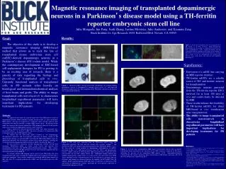
Magnetic resonance imaging of transplanted dopaminergic neurons in a Parkinson’s disease model using a TH-ferritin reporter embryonic stem cell lineJulie Mangada, Jun Peng, Junli Zhang, Justine Montoya,Julie Andersen, and Xianmin ZengBuck Institute for Age Research, 8001 Redwood Blvd, Novato, CA 94945 • Results: Goal: The objective of this study is to develop a magnetic resonance imaging (MRI)-based method that allows us to track the fate of transplanted mouse embryonic stem cell (mESC)-derived dopaminergic neurons in a Parkinson’s disease (PD) rodent model. While the contemporary development of ESC-based cell replacement therapies for PD is proving to be an exciting area of research, there is a paucity of data regarding the biology and physiology of transplanted cells in vivo. Currently, functional analysis of transplanted cells in PD animals relies heavily on histological and immunohistochemical analysis of host brains and grafts. The ability to image transplanted cells non-invasively to characterize longitudinal engraftment parameters will have important implications for developing treatments for PD patients. Methods: Our approach relies upon the targeted expression of the MRI reporter ferritin in dopaminergic neurons. While naturally occurring ferritin in cells is a source of intrinsic MRI contrast, cells that are engineered to ectopically express ferritin sequester more endogenous iron and thus provide a stronger MRI contrast agent 1. By placing ferritin under the control of the tyrosine hydroxylase (TH) promoter, relatively tissue-specific expression of ferritin in dopaminergic neurons can be achieved 2. Using a protocol of efficient derivation of mESC lines from transgenic mice described by Bryja 3, we derived a mESC line from a blastocyst of homozygote TH-ferritin male bred with C57BL/6.D2 F1 female. To validate that the TH-ferritin mESCs retained in vitro dopaminergic developmental potentials and tissue-specific expression of human ferritin in TH dopaminergic neurons, we generated TH+ cells from the TH-ferritin mESCs using two independent protocols. We first used the stromal cell co-culture method 4,5 and then the 5-stage protocol of dopaminergic differentiation of mESCs described by Kim et al6. We next wanted to determine whether dopaminergic neurons derived from transgenic TH-hFerritin mESC in culture could survive and function after transplantation into mice with MPTP-induced Parkinsonian lesions. Thus, neurons generated from coculture with PA6 stromal cells were stereotaxically implanted into the striata of mice that received MPTP. To evaluate whether MR imaging can be used to track TH-ferritin cells after transplant into brains, mice receiving the reporter grafts were sacrificed at both 4 and 12 weeks post transplant. After sacrifice, animals were perfused and the brains imaged via MR with both T2*-weighted gradient-echo (GRE) and T2-weighted spin-echo (SE) sequences. Figure 3. At the time of transplant, there was a high percent of TH+ neurons in the PA6 cocultures, though dopaminergic differentiation was not absolute as evidenced by the presence of GalC+ oligodendrocytes (A-B). Papain dissociation and low centrifugation did not compromise the integrity of the neurons as illustrated by the presence of long neuronal processes 24 hours after a cohort of cells for transplantation were replated onto adherent tissue culture dishes and observed with brightfield microscopy (C). • Significance: • Derivation of a mESC line carrying an MRI reporter (ferritin) • TH-ferritin mESCs are a reliable source of TH+ and ferritin+ neurons for transplantation • Dopaminergic neurons generated from the TH-ferritin reporter ESCs survived for at least 100 days in vivo and could clearly be detected by MRI • These results indicate the feasibility of TH-ferritin mESCs for direct MRI-based in vivo visualization after transplantation • The ability to image transplanted cells non-invasively to characterize longitudinal engraftment parameters will have important implications for developing treatments for PD patients • References: 1. Genove G, DeMarco U, Xu H, et al. A new transgene reporter for in vivo magnetic resonance imaging. Nat Med. 2005;11:450-454. 2. Kaur D, Yantiri F, Rajagopalan S, et al. Genetic or pharmacological iron chelation prevents MPTP-induced neurotoxicity in vivo: a novel therapy for Parkinson's disease. Neuron. 2003;37:899-909. 3. Bryja V, Bonilla S, Cajanek L, et al. An efficient method for the derivation of mouse embryonic stem cells. Stem Cells. 2006;24:844-849. 4. Kawasaki H, Mizuseki K, Nishikawa S, et al. Induction of midbrain dopaminergic neurons from ES cells by stromal cell-derived inducing activity. Neuron. 2000;28:31-40. 5. Zeng X, Chen J, Sanchez JF, et al. Stable expression of hrGFP by mouse embryonic stem cells: promoter activity in the undifferentiated state and during dopaminergic neural differentiation. Stem Cells. 2003;21:647-653. 6. Kim JH, Auerbach JM, Rodriguez-Gomez JA, et al. Dopamine neurons derived from embryonic stem cells function in an animal model of Parkinson's disease. Nature. 2002;418:50-56. • Figure 1. TH-ferritin mESCs showed typical ESC morphology (A) and high alkaline phosphatase activity as determined by enzyme activity assay (C), and expressed pluripotent marker Oct4 (D). The integration of the TH-ferritin construct in the genome was verified by RT-PCR (B). Figure 2.TH+ cells were generated from the TH-ferritin mESCs using two independent protocols. We first used the stromal cell co-culture method 4,5, and showed that TH+ cells could be efficiently generated from TH-ferritin mESCs (A-C). We then used the 5-stage protocol of dopaminergic differentiation of mESCs described by Kim et al6 and generated neural stem cells (NSCs) and dopaminergic neurons. mESC cultured via embryoid body (EB) formation lead to a high percent of cells positive for the NSC marker Nestin (D). Additional staining showed that midbrain dopaminergic marker Nurr1 was expressed in cells after removal of FGF2 and an additional 20 days of differentiation (E-G). No difference in neuronal inducing efficiency and timing was observed between the reporter and WT mESC lines. In addition, double immunostaining of TH and ferritn revealed the co-expression of TH and ferritin in dopaminergic neurons, indicating the tissue-specific expression of ferritin in the transgenic cells (H). Figure 4 12 weeks post transplantation MRI images reveal robust contrast only in reporter-transplanted brains proximal to the injection site in GRE sequences compared to SE images, indicating the presence of the transplanted MRI reporter (A-D). Immunohistochemistry confirmed that this region contained a high concentration of TH-ferritin expressing cells (E-G) that were absent in the control brain (H-J).