Download
1 / 46
480 likes | 698 Vues
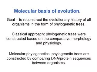
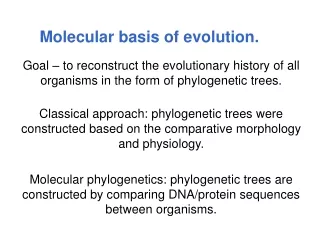
Molecular Basis of Lung Disease. Exons. Polyadenylation signal. Start of transcription. Termination codon UAA UAG UGA. Initiation codon ATG. Introns. 5’ untranslated region. 3’ untranslated region. Basic Gene Structure. Promoter. HISTORY AND PHYSICAL FINDINGS.

E N D
Exons Polyadenylation signal Start of transcription Termination codon UAA UAG UGA Initiation codon ATG Introns 5’ untranslated region 3’ untranslated region Basic Gene Structure Promoter
HISTORY AND PHYSICAL FINDINGS • J.B., a 2-year-old boy, was referred to the pediatric clinic for evaluation of poor growth • During infancy, J.B. had diarrhea and colic that resolved when an elemental formula was substituted for his standard formula. As table foods were added to his diet, he developed malodorous stools containing undigested food particles • During his second year, J.B. grew poorly, developed a chronic cough, and had frequent upper respiratory infections. No one else in the family had poor growth, feeding disorders, or pulmonary illnesses. • On physical examination, J.B.'s weight and height plotted less than the 3rd percentile and his head circumference at the 10th percentile. He had a severe diaper rash, diffuse rhonchi, and mild clubbing of his digits • the pediatrician requested several tests, including a test for sweat chloride concentration by pilocarpine iontophoresis; the sweat chloride level was 75 mmol/L (normal, <40 mmol/L; indeterminate, 40 to 60 mmol/L), a level consistent with cystic fibrosis. • On the basis of this result and the clinical course, the pediatrician diagnosed J.B.'s condition as cystic fibrosis. J.B. and his parents were referred to the cystic fibrosis clinic for further counseling, mutation testing, and treatment.
CFTR:CF transmembrane regulator (positional cloning) • Since the 1960s, cystic fibrosis (CF) has been one of the most publicly visible of all human monogenic diseases • It is the most common fatal autosomal recessive genetic disorder of children in white populations, with an incidence of approximately 1 in 2500 white births and a carrier frequency of about 1 in 25 • The gene cloned by positional cloning in 1989 • Shortly after the CF gene was cloned, physiological analyses demonstrated that the protein encoded by the CFTR gene is a regulated chloride channel located in the apical membrane of the epithelial cells affected by the disease.
Autosomal Recessive Pedigree illustrating recessive inheritance Autosomal recessive disease occurs only in individuals with 2 mutant copies, which they inherited from their normal heterozygous parents (barring the rare new mutation)>
Genotype: A A (Homozygous) Genotype: A B (Heterozygous) A B A A Gene Chromosome 6 Maternal copy Chromosome 6 Paternal copy DNA Basic terminology Single gene disorder - determined by the alleles at a single locus
“Salty Baby Syndrome" Mechanism Underlying Elevated Sodium Chloride Levels in the Sweat of Patients with Cystic Fibrosis A major pathway for Cl– absorption is through CFTR: The result is that total sodium chloride flux is markedly decreased, leading to increased salt content
CFTR is expressed in surface epithelium and serous cells at the base of submucosal glands in a porcine lung sample
Submucosal glands of a patient with cystic fibrosis are filled with mucus, and mucopurulent debris Pseudomonas aeruginosa A median cross section of a lung from a patient with CF
CFTR gene mapped to chromosome 7q31 Spans about 190 kb of DNA 27 exons, is predicted to encode a large integral membrane protein of about 170 kD Has five domains: two membrane-spanning domains, each with six transmembrane sequences; two nucleotide (ATP)-binding domains; and a regulatory domain with multiple phosphorylation sites TheCFTRGene and Protein .
no chloride-channel function lead to misfolded CFTRprotein that is prematurely degraded
0 50 100 150 200kb 24 11 19 14b 16 4 5 9 13 18 20 22 2 3 10 21 1 7 23 6a 14a 8 12 15 17a 6b 17b Recommended mutations to be screen in CFTR gene TMD1 TMD2 NBD1 NBD1 R domain A455E R117H R117C W1282X S1255X N1303K W1316X R33W R347C R347P G542X R553X G493X G551D G551S R1066H R1066C R1066L F508 I507 3849+10kbC>T
A total of 33 mutations had been screen including the recommended 25 in our patients using OLA. G85E 394delTT 621+1G>T 1898+1G>A 3849+10kbC>T 711+1G>T 2183AA>G I148T 621+1G>T R117H A455E 3905insT W1282X 2789+5G>A 1717-1G>A 2184delA 24 11 19 14b 16 4 5 9 13 18 20 22 2 3 10 21 1 7 23 8 12 15 R347H R347P R334W 1078delT V520F I507del Δ F508 S549R S549N G542X G551D R553X R560T N1303K 3876delA 3120+1G>A 3659delC R1162X
OLA Principle OLA with mutant DNA OLA withnormal DNA G A normal mutant Fluorescent dye Fluorescent dye mutant common PO4 normal PO4 common A G C T mutant DNA target normal DNA target ligation ligation G A Normal OLA product mutant-OLA product Is slightly longer than normal OLA product due to longer mobility modifier Target Specific Probe With Mobility Modifier Result on 3100: normal heterozygous homozygous mutant
Example on Oligonucletide Ligation Assay Results ∆F508/∆F508 ∆F508/∆F508 ∆F508 ∆F508/- ∆F508 G85E ∆F508/G85E
Allelic frequencies of mutations in the Jordanian CF patients 10.3% 3.9% 5.2% G85E W1282X 711+1G>T 24 11 19 14b 16 4 5 9 13 18 20 22 2 3 10 21 1 7 23 8 12 15 Δ F508 N1303K 24.6% 1.3%
CFTR gene sequencing is the choice 77 patients with sweat chloride value more than 60 mmol/L 30 tested –ve for the 33 mutations 47 tested +ve for the 33 mutations Heterozygous (24) Compound Heterozygous (5) Homozygous (18)
SERPINOPATHIES Serine Proteinase Inhibitors α1-antitrypsin
Alpha1-Antitrypsin Deficiency and Conformational Instability Pathologicpolymerization of the variant alpha1-antitrypsin before itssecretion from hepatocyte.
Mechanism of Inhibition of Proteases by Serpins neutrophil elastase
The main function of α1-antitrypsin is toprotect the tissues, and especially the elastic tissue ofthe lungs, against the enzyme neutrophil elastase
Z allele • One in 10 people of European descent is a carrier of one of two mutations in alpha1-antitrypsin that result in a partial deficiency of the inhibitor • S mutation (Glu264Val), which in homozygotes results in a 40 percent decrease in plasma alpha1-antitrypsin concentrations. This by itself poses a negligible threat to health, • but the S variant becomes important if it is coinherited with the more severe Z mutation (Glu342Lys). Some 4 percent of Northern Europeans (3 percent in the United States) carry the Z mutation; in homozygotes, this results in an 85 percent deficit in plasma alpha1-antitrypsin concentrations • Consequently, the plasma concentrations of alpha1-antitrypsin in both ZZ homozygotes and SZ compound heterozygotes are insufficient to ensure lifetime protection of the lungs from proteolytic damage, especially in smokers.
The effect of smoking on the survival of patients with α1-antitrypsin deficiency
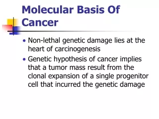
Cancer as a genetic disease • whether sporadic or familial, cancer is fundamentally due to mutation in various genes controlling cell growth or cell death • once initiated, the cancer evolves by accumulating additional mutations in other genes • leads to an ever-worsening cascade of mutations • Original clone of neoplastic cells can evolve into numerous sublineages with different but overlapping mutations Tumor suppressor gene Protooncogene
Classifying the genes involved in cancer • Oncogenes – mutant forms of genes (proto-oncogenes) that positively regulate cell proliferation and cell survival -usu dominant, gain-of-fn mutations • Tumor suppressors – genes which function to block tumor development by negatively regulating cellular growth-usu need loss of both copies • Cellular maintenance genes – responsible for the detection and repair of genetic damage in cells
Receptor tyrosine kinases -transduce an extracellular signal inward -bind a ligand, conformational change that results in kinase activity, leading to phosphor- lation of cellular proteins -pt mut’s cause receptors to be constitutively active RET mut-multiple endocrine neoplasia MET mut-hereditary papillary renal carcinoma RET, MET Ras, Abl Myc
RAS family of proto-oncogenes • One of the first activated oncogenes discovered by the DNA transformation assay • Encodes a small guanosine triphosphate (GTP) –binding protein (G-protein) • 3 members of this family; H-RAS, K-RAS, N-RAS • Serves as an “on/off” switch to activate or inhibit downstream molecules when bound to GTP • The protein’s effect is ended by self-directed cleavage of GTP
RAS family of proto-oncogenes • Ras associates with the plasma membrane • Ras relays signals from the cell surface receptors to the nucleus, functioning as a switch • ‘Active’ when GTP is bound • ‘Inactive when the hydrolyzed GDP is bound
RAS oncogene activation by nucleotide substitution • Conversion to oncogene usually due to a point mutation in the gene where: • the ras protein is able to signal continuously, even in absence of GTP • the ras protein is unable to hydrolyze GTP to turn “off” the signal • Leads to the continuous activation of multiple downstream signaling pathways inducing cell proliferation • Mutations in the 3 RAS genes are found in 10-15% of all human cancers
RAS mutation in human cancers • H-RAS mutated in 10% of all bladder cancer • K-RAS mutations in about 50% of colorectal cancers, 70-90% of pancreatic cancers and 30% of lung adenocarcinomas as well as in ovarian, breast skin liver and kidney • N-RAS mutations have been detected in 20-30% of acute nonlymphocytic leukemias
Codon 12 G-T The BRAF–mitogen-activated and extracellular-signal regulated kinase kinase (MEK) –extracellular signal-regulated kinase (ERK) cascade often determines proliferation and becomes deregulated in certain cancers
EGFRin lung adenocarcinomain approximately 10% of specimens from patients in the UnitedStates and in 30 to 50% of specimens from patients in Asia. The mutations occur with increased frequency inwomen and nonsmokers
The two-hit hypothesis • Tumorigenesis requires loss of function of both copies of tumor suppressor • At clinical level – dominant inheritance (looks like only a single mutation is required) • At cellular level – two mutations required for tumor to develop • First hit may be inherited or occur somatically • Second hit occurs somatically • One mechanism – loss of heterozygosity - LOH: individual is heterozygous in normal tissues at a specific marker but tumor cells contain only 1 of the 2 alleles
Five-gene signature is closely associated with relapse-free and overall survivalamong patients with NSCLC • NEJM 356;1 www.nejm.org january 4, 2007 • DUSP6, MMD,STAT1, ERBB3, and LCK
Examples of microRNA profiles in human solid and liquid cancers
MiRNA profiling as a prognostic tool • The expression of both miR-155 (high levels) and let-7a-2 (low levels) has been shown to correlate with poor survival in 104 United States patients with lung cancer. • In an independent study of 143 Japanese patients with lung cancer, reduced let-7 expression was found to significantly correlate with a shorter survival time after potentially curative surgery • Ras overexpression was associated with a reduced survival time. As let-7 negatively regulates Ras, these results show the existence of a link between let-7, Ras expression and the life expectancy of patients with lung cancer.