Infrared Spectroscopy
Infrared Spectroscopy. The structure of new compounds that are isolated from natural sources or prepared in the lab must be determined (and/or verified). Chemical analysis Spectroscopy Spectroscopic techniques are non-destructive and generally require small amounts of sample.

Infrared Spectroscopy
E N D
Presentation Transcript
Infrared Spectroscopy • The structure of new compounds that are isolated from natural sources or prepared in the lab must be determined (and/or verified). • Chemical analysis • Spectroscopy • Spectroscopic techniques are non-destructive and generally require small amounts of sample
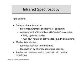
Infrared Spectroscopy • Four common spectroscopic techniques used to determine structure: • Infrared Spectroscopy (IR) • Mass Spectrometry (MS or Mass Spec) • Nuclear Magnetic Resonance Spectroscopy (NMR) • Ultraviolet Spectroscopy
Infrared Spectroscopy • Infrared spectroscopy: • Used to determine the functional groups present (or absent) in a molecule http://riodb01.ibase.aist.go.jp/sdbs/ (National Institute of Advanced Industrial Science and Technology, 1/5/11)
Infrared Spectroscopy • Infrared spectroscopy is a type of absorption spectroscopy: • Any technique that measures the amount of light absorbed by (or transmitted through) a compound as a function of the wavelength of light • Sample is irradiated by a light source • Amount of light transmitted (or absorbed) at various wavelengths is measured by a detector • A spectrum is obtained. • Graph of light transmitted (or absorbed) as a function of wavelength
Infrared Spectroscopy • Infrared spectroscopy uses light from the infrared region of the electromagnetic spectrum. • The absorption of IR radiation leads to absorption bands in the IR spectrum. • The position of a band is reported in wavenumbers (u ) • the number of wavelengths per cm • u = 10,000 where l = mm l • Directly proportional to energy
Infrared Spectroscopy • The atoms in a molecule are in constant motion. • The covalent bond between two atoms acts like a spring, allowing the atoms to vibrate (stretch and bend) relative to each other.
Infrared Spectroscopy • The absorption of IR radiation increases the amplitude of the various types of bond vibrations. • Stretching • Symmetric • Asymmetric • Bending
Infrared Spectroscopy • Since energy is quantized, covalent bonds can vibrate/stretch only at certain allowed frequencies. • The position of an absorption band correlates with the type of chemical bond.
Infrared Spectroscopy • The frequency of an absorption band in an IR spectrum depends primarily on: • Type of vibration • Stretching vibrations: higher frequency • Bending vibrations: lower frequency • Masses of the atoms in a bond • Strength of the bond or bond order
Infrared Spectroscopy • The polarity of a bond has a significant impact on the intensity of an IR absorption band. • Vibrations that cause a significant change in the dipole moment of a chemical bond lead to strong absorption bands. • Vibrations that result in no change/very little change in dipole moment lead to very weak or no absorption band. • Symmetrical bonds often exhibit very weak or no absorption band.
Infrared Spectroscopy • Each molecule has a unique IR spectrum. • The IR spectrum is a “fingerprint” for the molecule. • IR spectrum results from a combination of all possible stretching and/or bending vibrations of the individual bonds and the whole molecule. • Simple stretching: ~1600-4000 cm-1. • Complex vibrations: 600-1400 cm-1, called the “fingerprint region.”
Infrared Spectroscopy • IR Spectrum of n-octane
Infrared Spectroscopy • An IR spectrum is used to identify functional groups that are present (or absent). • Cannot conclusively identify a structure by IR alone unless an IR spectrum of an authentic (known) sample of the compound is available. • Absorptions from specific functional groups are found in certain regions of the IR spectrum.
Carbon-Carbon Bonds • Increasing bond order leads to higher frequencies: • C-C 1200 cm-1(fingerprint region) • C=C 1600 - 1680 cm-1 • CC 2200 cm-1 (weak or absent if internal) • Conjugation lowers the frequency: • isolated C=C 1640-1680 cm-1 • conjugated C=C 1620-1640 cm-1 • aromatic C=C approx. 1600 cm-1 • C=C peaks are generally weak to moderate in intensity.
Carbon-Hydrogen Bonds • Bonds with more s character absorb at a higher frequency. • sp3 (alkane) C-H • just below 3000 cm-1 (to the right) • sp2 (alkene or aromatic hydrocarbon) C-H • just above 3000 cm-1 (to the left) • sp (alkyne) C-H • at 3300 cm-1
O-H and N-H Bonds • Both O-H and N-H stretches appear around 3300 cm-1, but they look different. • Alcohol O-H • broad with rounded tip when hydrogen bonding is present (sharp in the absence of hydrogen bonding) • Secondary amine (R2NH) • Broad (usually) with one sharp spike • Primary amine (RNH2) • Broad (usually) with two sharp spikes. • No signal for a tertiary amine (R3N)
NH Bend • A broad, round peak may be observed around 1600 cm-1 for the N – H bend, especially with primary amines. NH2 stretch N-H bend N-H bend has a different shape than an aromatic ring or C=C
Carbonyls • Carbonyl stretches are generally strong: • Aldehyde ~1710 cm-1 • Ketone ~1710 cm-1 • Carboxylic acid ~1710 cm-1 • Ester ~1730 - 1740 cm-1 • Amide ~1640-1680 cm-1 • Conjugation shifts all carbonyls to lower frequencies. • Ring strain shifts carbonyls to higher frequencies.
1743 1245 Esters • C=O stretch at ~ 1730-1740 cm-1 and • C-O stretch at 1000-1300 cm-1 (broad) (Note: other functional groups may have peaks in the 1000-1300 cm-1 region too!) strong
Amides • C=O stretch at 1640-1680 cm-1(sometimes a double peak) • N-H stretch (if 1o or 2o) around 3300 cm-1
Nitriles • C N absorbs just above 2200 cm-1 (med – strong) • The alkyne C C signal is much weaker and is just below 2200 cm-1
IR Spectroscopy Example: Interpret the following IR spectrum by assigning each of the major peaks. Identify what functional group(s) are present. 3403 cm-1 1604 cm-1 http://riodb01.ibase.aist.go.jp/sdbs/ (National Institute of Advanced Industrial Science and Technology, 12/30/09)
IR Spectroscopy Example: Interpret the following IR spectrum by assigning each of the major peaks. Identify what functional group(s) are present. 2733 cm-1 2814 cm-1 1642 cm-1 1691 cm-1 http://riodb01.ibase.aist.go.jp/sdbs/ (National Institute of Advanced Industrial Science and Technology, 12/30/09)
1721 Infrared Spectroscopy Example: Which one of the following compounds is the most reasonable structure for the IR spectrum shown below? http://riodb01.ibase.aist.go.jp/sdbs/ (National Institute of Advanced Industrial Science and Technology, 12/30/09)
1603 1689 Infrared Spectroscopy Example: Which of the following compounds is the most reasonable structure for the IR spectrum shown below? http://riodb01.ibase.aist.go.jp/sdbs/ (National Institute of Advanced Industrial Science and Technology, 12/30/09)