Levels of Analysis
380 likes | 521 Vues
Explore the intricacies of cortical lobes involved in vision, audition, and language. This overview delves into the "what vs. where" pathways and networks critical to understanding visual perception. Key concepts include retinotopy, the spatial mapping of visual fields, and the hierarchical and functional organization of visual areas. We examine cortical computations, motion detection, color processing, and facial recognition. Discover the implications of retinotopic maps for cognitive functions such as attention and visual imagery, and learn how low-level visual processing builds towards more complex representations.

Levels of Analysis
E N D
Presentation Transcript
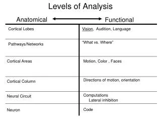
Cortical Lobes Vision, Audition, Language “What vs. Where” Pathways/Networks Cortical Areas Motion, Color , Faces Directions of motion, orientation Cortical Column Computations Lateral inhibition Neural Circuit Neuron Code Levels of Analysis Anatomical Functional
Organizational principles of the visual system • Retinotopy • Anatomical organization • “Hierarchical” organization • Functional organization based on neural response properties
Retinotopy • Retinotopy = a spatial map of the visual field with retina-based coordinates • Implication = a spot of light in the visual field will activate a ‘spot’ of cortex based on eye position
Retinotopy sets the stage • Retinotopy = a spatial map of the visual field with retina-based coordinates • Implication = a spot of light in the visual field will activate a ‘spot’ of cortex based on eye position • Retinotopicmaps define multiple visual areas (i.e., each “area” has its own map of the visual field)
Macaque Retinotopy Source: Tootell et al., 1982
V2d LVM V2v UVM Polar angle UVM Eccentricity HM LVM V1 F
V2d LVM V2v UVM LVM V1 UVM F HM
V2d LVM V2v UVM LVM V1 UVM F HM
8 Hz flicker (checks reverse contrast 8X/sec) • good stimulus for driving visual areas • subjects must maintain fixation (on center dot) EXPANDING RINGS ROTATING WEDGES
Spatial Maps Beyond Visual Cortex • Hagler, D. and M.I. Sereno (2005) Spatial maps in frontal and prefrontal cortex. Neuroimage29:567-577 • Sereno, M.I., S. Pitzalis, and A. Martinez (2001) Mapping of contralateral space in retinotopic coordinates by a parietal cortical area in humans. Science294:1350-1354.
MIDTERM • Worth 99% of final grade
Retinotopy: a powerful tool for studying the mind • Retinotopy & Mental Imagery • Retinotopy & Attention • Retinotopy & Spatial Memory
Visual Imagery (old days) • Does visual imagery rely on symbolic (“language-like”) processing or depictive (picture-like) representations? (Pylyshyn vs. Kosslyn) • Use RT in standard cognitive psychology paradigm to infer “visual-like” behavior.
Visual Imagery (old days) RT depends on position of probe Imagery a ‘sequential’, ‘depictive’ operation x f
Retinotopy & Visual Imagery • Slotnick SD, Thompson WL, Kosslyn SM. Visual mental imagery induces retinotopically organized activation of early visual areas. Cereb Cortex. 2005 Oct;15(10):1570-83. • Does imagery evoke cortical activity with precise visual field topography?
Perception Imagery Attention
Organizational principles of the visual system • Retinotopy • Anatomical Organization • “Hierarchical” organization • Functional Organization based on neural response properties
Functional “Hierarchy” Properties of lower areas (lower = anatomically closer to the retina) form the building blocks of higher areas. • The visual feature(s) that elicit an increase in spike rate becomes progressively more complex. • “Selectivity” • “Tuning” • The area of visual space that elicits an increase in spike rate in a neuron becomes progressively larger. • “Receptive field”
Input Image Early visual cortex Higher visual cortex Small & Simple Large & Complex
Functional “Hierarchy” Properties of lower areas (lower = anatomically closer to the retina) form the building blocks of higher areas. • The visual feature(s) that elicit an increase in spike rate becomes progressively more complex. • “Selectivity” • “Tuning” • The area of visual space that elicits an increase in spike rate in a neuron becomes progressively larger. • “Receptive field”
LGN V1 Connections • More “complex” tuning/selectivity • Larger receptive fields
LGN Response Properties • Selectivity = light/dark spots of light • Receptive fields = are extremely small. Fovea approximately few minutes (60 minutes = 1 degree)
p.156 LGN V1
Functional “Hierarchy” • The visual feature(s) that elicit an increase in spike rate becomes progressively more complex. • “Selectivity” • “Tuning” • The area of visual space that elicits an increase in spike rate in a neuron becomes progressively larger. • “Receptive field”
Movie Time On-center Off-center Direction Cell Simple Cell
Functional “Hierarchy” • The visual feature(s) that elicit an increase in spike rate becomes progressively more complex. • “Selectivity” • “Tuning” • “Selective” = the type of stimulus that a neuron responds to • “Tuning” = a continuous stimulus dimension • A neuron can be “selective” (e.g., a “face cell”) but that doesn’t necessarily imply “tuning”
2 3 10 4 Can “Tuning” be measured with fMRI? 0 1
Other forms of “tuning” • Other visual examples • Other auditory examples • …
Beyond V1 • Assumption that the Hubel & Wiesel approach will extend to other visual areas • “Simple” feature selectivity combines to form more “complex” feature selectivity • “Hierarchical Feedforward Model” • Observations in extra-striate areas • Selectivity becomes more complex • Receptive fields are larger
Next week Thursday