THE HEARTBEAT
THE HEARTBEAT. HEART ANATOMY. Heart Physiology. The heart rate is the number of times the heart beats per minute. Regulated by the autonomic nerve supply, sympathetic fibers to the heart, located in the superior, middle and inferior cardiac nerves.

THE HEARTBEAT
E N D
Presentation Transcript
THE HEARTBEAT HEART ANATOMY
Heart Physiology • The heart rate is the number of times the heart beats per minute. • Regulated by the autonomic nerve supply, sympathetic fibers to the heart, located in the superior, middle and inferior cardiac nerves. • Impulses traveling over them insure that the heart beats fast enough to maintain good circulation during any activity.
Parasympathetic fibers, traveling in the vagus nerves, tend to slow the heart, and serve as cardioinhibitor nerves. • This insures the heart does not beat too fast or too slow when the individual is resting or being active. • The heart is composed of a fused mass of contractile cell, and obeys the all-or-nothing law of conductivity.
Under any conditions, the heart, when stimulated will contract maximally or not at all. That is not good!! • The heart cannot tetanize ( a sustained contraction due to repeated stimulus, such as skeletal muscle). • The heart has an extra long refractory period (0.3 sec. = 65 times as long as a skeletal muscle.
Stimuli applied during systole will not produce an additional response. • An extrasystole can be demonstrated. • These are premature beats and are always followed by a long compensatory pause.
The Cardiac Cycle • Events that occur during one heartbeat, or contraction and subsequent relaxation. • Average of 72 beats per minute. • Must be completed in 0.8 sec. • The amount of blood pumped is about 70 cc of blood per stroke. This is called stroke volume
Figuring Stroke Volume • Cardiac Output = Heart Rate x Stroke Vol. • Heart Rate of 70 x Stroke Volume of 70 = 4900 cc of blood which is normal base line. • 5 liters of blood are pumped each minute by the heart when physical activity is no more strenuous than sitting in a chair. • Loss of blood, shock, cardiogenic shock, etc.
ALL OF THESE FACTORS AFFECT THE HEART !! • THE END
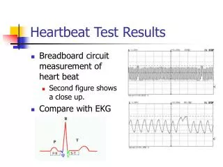
HOW DOES THE HEART BEAT? • Two general classes of cardiac muscle cells involved in the normal heartbeat: contractile cells and the conducting system. • Cardiac muscle cells have a long refractory period (resting). • Rapid stimulation produces isolated rather than tetanic (abnormally prolonged contractions that results from disturbances in the electrolyte balance) contractions.
Action Potential of Cardiac Cells • Rapid Depolarization: Sodium channels open, and the membrane suddenly becomes permeable to sodium. Causes rapid depolarization. The channels are called fast channels because they are open only a few milliseconds. • The Plateau: As sodium channels close, calcium channels open. The channels are called slow channels because they open slowly and remain open for a longer period (175 milliseconds). • Repolarization: As the plateau continues, calcium channels begin closing and slow potassium channels begin opening. Causes rapid repolarization and resting potential.
Refractory Period • For sometime after an action potential begins, the membrane will not respond to a second stimulus. • This period is called the refractory period. • Absolute refractory period – The membrane cannot respond at all. • Relative refractory period – Sodium channels are closed but capable of opening.
Plateau Rapid depolarization Repolarization
That is why you need to have dairy products, small amounts of salt, and fresh fruits and vegetables in your diet. Your heart depends on calcium, sodium, and potassium to cause a heartbeat !!
Conducting System • Cardiac muscle contracts on its own, in absence of neural or hormonal stimulation. • This is called automaticity. • The conducting system is a network of specialized cardiac cells that initiates and distributes electrical impulses.
The conduction system consists of the following: Sinoatrial (SA) node, located in the wall of the right atrium. Atrioventricular (AV) node, located at the junction between the atria and ventricles. Conducting cells, which interconnect the two nodes and distribute the impulse throughout the myocardium. The AV bundle, bundle branches, and Purkinje fibers, which distribute the stimulation to the ventricular myocardium.
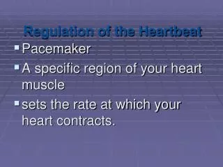
Sinoatrial Node • Embedded in the posterior wall of the right atrium near the superior vena cava. • Contains pacemaker cells that establish heart rate. • SA node is also known as the cardiac pacemaker. • The contractile stimulus is passed to the right and left atria.
Atrioventricular Node • Sits within the floor of the right atrium • The rate of propagation impulse slows as it leaves the internodal pathways and enters the AV node. • This delay is important because the atria must contract before the ventricles. • When the atrial completes its contraction the ventricular contraction begins.
Bundle of His, Bundle Branches, & Purkinje Fibers • The AV node and Bundle of His is the only electrical connection between the atria and ventricles. • The impulse travels to the right and left bundle branches. • The left branch supplies the left ventricle and the right branch supplies the right ventricle.
Both branches extend toward the apex and fan out beneath the endocardial surface. • The Purkinje fibers relay action potentials to the ventricles. • Ectopic pacemakers bypass the conducting system disrupting the timing of the contraction.
Signal 2, contraction of the atria
Impulse to the AV node
Impulse travels down the bundle of His, to the right and left bundle branches