

THE HEART:
E N D
Presentation Transcript
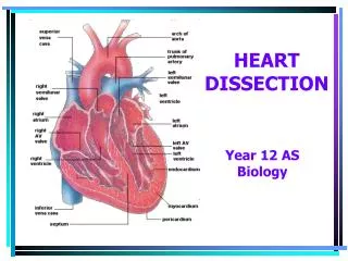
THE HEART: The heart is a hollow, fibromuscular, thick-walled organ located in the middle mediastinum of the thorax. It is also a double, self-adjusting muscular pump, which receives oxygenated blood from the pulmonary circulation and propels blood through the systemic circulation to all the tissues of the body. A little bit larger than the closed fist of the owner, it has an average weight range of 280 – 340gm (male) and 230 – 280gm (female). It is conical in shape and possesses a base, three surfaces and an apex as follows:
Base (Vertebral or Posterior surface): This is directed posterosuperiorly and to the right, and lies opposite T5-T8. It is formed by the two atria. Diaphragmatic (Inferior) Surface: This surface is directed inferiorly and related to the central tendon of the diaphragm. It is also formed by the two ventricles. Sternocostal (Anterior) Surface: This surface lies directly behind the sternum. It is formed by the two ventricles.
Pulmonary (Left) Surface: This is the surface in contact with the left lung. It is formed by the left ventricle. Apex: The apex of the heart is formed by the left ventricle and directed inferolaterally to the left. It is located in the left 5th intercostal space about 9cm from the midline and below the left nipple.
SURFACE ANATOMY OF THE HEART In actual clinical practice, the borders of the sternocostal (Anterior) surface are used to describe the surface anatomy of the heart. This surface has four borders, viz. The superior border which is formed by the right and left auricles and the pulmonary trunk The left border which is formed by the left ventricle and the left auricle The right border which is formed by the right atrium The inferior border which is formed by the right and left ventricles. The projection of these borders to the chest wall is illustrated in the diagram below (Fig. ***) By the letters ABCD
AB:This represents the upper border. BC:This represents the left border of the heart. CD:This represents the inferior border of the heart. DA:This represents the right border of the heart. The area of the chest wall which maps out the surface projection of the heart is referred to as the Precordium.Drugs could be administered into the heart in this area in situations of emergency as in Cardiopulmonary Resuscitation (CPR). This is also the area at which cardiac massage is administered.
OTHER SURFACE FEATURES OF THE HEART The Coronary Sulcus (Groove):This is the surface projection of the atrioventricular septum. The Interventricular sulcus (Groove):This is the surface projection of the interventricular septum. The Sulcus Terminalis: This is the shallow vertical groove on the posterior aspect of the right atrium It separates the two different embryological derivatives of the right atrium. Deep to this groove in the cavity of the right atrium lays the cristal terminalis.
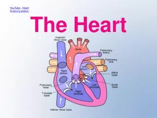
THE CAVITIES OF THE HEART The heart is compartmentalized into two groups of chambers, the atria and the ventricles. While the atria are separated by non-perforated membranous septum and the ventricles by non -perforated musculomembranous septum, the atria are separated from the ventricles by valves (Atrioventricular valves). The right atrium and ventricle form the right pump while the left pump is formed by the left atrium and ventricle
THE RIGHT ATRIUM: • This cavity receives deoxygenated blood from the systemic circulation through the superior and inferior vena cava. • Deoxygenated blood from the heart wall is also drained into this cavity via the coronary sinus. • When the right atrium contracts, it pumps deoxygenated blood in it into the right ventricle via the right atrioventricular (tricuspid) valve. The capacity of the cavity is increased by the muscular pouch called the right auricle
THE RIGHT VENTRICLE • The right ventricle receives deoxygenated blood from the right atrium via the tricuspid valve. • It has a thicker wall than the right atrium which enables it to pump deoxygenated blood to the pulmonary circulation via the pulmonary trunk. • A semilunar valve is located between this ventricle and the pulmonary trunk
THE LEFT ATRIUM • The left atrium forms 2/3 of the base of the heart. • It also possesses an auricle and receives oxygenated blood from the right and left lungs through four pulmonary veins
THE LEFT VENTRICLE • This ventricle forms the entire pulmonary surface, the entire apex, and contributes to the anterior and diaphragmatic surfaces of the heart. • The wall is twice as thick as the right ventricular wall except at the apex. • It receives oxygenated blood from the left atrium via the bicuspid (Mitral) valve and pumps the blood into the systemic circulation via the ascending aorta. • There is a valve at the opening of the ventricle into the aorta (Semilunar valve)