CYCAS Structure, Reproduction & Life-Cycle
5.23k likes | 39.56k Vues
CYCAS Structure, Reproduction & Life-Cycle. Dr. Maninder Kaur Associate Professor Botany Post Graduate Government College for Girls Sector-11, Chandigarh. Systematic Position. Gymnospermae Division: CYCADOPHYTA Class: CYCADOPSIDA Order: CYCADALES Family: CYCADACEAE Genus: CYCAS

CYCAS Structure, Reproduction & Life-Cycle
E N D
Presentation Transcript
CYCASStructure, Reproduction & Life-Cycle Dr. ManinderKaur Associate Professor Botany Post Graduate Government College for Girls Sector-11, Chandigarh
Systematic Position • Gymnospermae • Division: CYCADOPHYTA • Class: CYCADOPSIDA • Order: CYCADALES • Family: CYCADACEAE • Genus: CYCAS (Greek word Kycas = Cocopalm)
Distribution & Occurrence • Includes 20 Species • Occurs wild or cultivated in tropical and sub-tropical regions • South of Eastern Hemisphere • e.g. S. Japan, India, China, N. Australia, E. Coasts of Africa, Myanmar, Bangladesh, Mauritius, Nepal, etc. • 6 species Indian – 4 wild & 2 cultivated • C circinalis, rumphii, pectinata & beddomei • C. revoluta & C. siamensis
Sporophytic Plant Body • Plants are low and palm-like, height 4-8 feet • Tallest species, C. media – upto 20 feet high • Stem unbranched, columnar and covered with persistent leaf bases • Leaf segment remains circinnatelyinvolute within the bud – leaves dimorphic • Female reproductive structures – the megasporophylls are not aggregated in cones • Ovules (2 or more) borne on the lower margins in ascending order
External Morphology • Stem – Cycas plant shows tuberous stem when young, becoming columnar and unbranched later • Leaf – Shoot apex is protected by a rosette of brown scale leaves • Plant grows very slowly adding a new crown of leaves every 1 or 2 years, alternating with crown of scale leaves
External Morphology • The pinnately compound megaphyllous leaves have 80-100 pairs of leaflets arranged on the rachis • Circinnateptyxis of young leaves is a fern like character • Leaf base is rhomboidal in shape and attaches the leaf transversely to the stem • The leaflets are thick , leathery in texture, ovate or lanceolatein shape & photosynthetic in function.
External Morphology • Scale leaves are very small, rough and dry, triangular in shape and brown in colour, thickly coated in ramenta • Root is of two types-normal and coralloid. • Normal tap-roots grow from the radicle deep inside the soil giving out lateral branches • Some of the lateral roots grow apogeotropically towards the surface of soil and branch dichotomously • These roots are short, thick and swollen at the tips. Cluster of coralloid roots
External Morphology • The much branched mass appears like a coral on the soil surface hence called coralloid roots • Do not bear root caps • The cluster has lenticel like apertures • Become infested by N2 fixed blue-green algae (cyanobacteria); bacteria & diatoms e.g. Nostocpunctiforme, Anabaena cycadacaerum • Symbiotic relationship thus established
Anatomy Root • Young root shows typical structure like that of a dicotyledonous root • Outermost layer, epiblema, encloses the parenchymatous cortex interspersed with tannin cells and mucilage canals • Endodermis with casparian thickenings • Pericycle is multilayered with thin cells having starch grains • Vascular tissue within is typically radial • Roots usually diarch to tetraarch, rarely polyarch • Vessels absent in vascular tissue • Pith reduced or absent
Anatomy – Root • Older roots show secondary growth • Cambial ring is initiated between xylem & phloem and completed by differentiation in inner layer of pericycle adjacent to protoxlem elements • These cambial cells are meristematic and add secondary xylem on the inside and secondary phloem towards cortex • Alongside phellogen (cork cambuim) develops in outermost layer of cortex below the epidermis • This produces dead cork cells (phellem) towards outer side and living secondary cortex cells (phelloderm) on the inside. • Lenticels are developed in old roots
Anatomy – Root Coralloid Roots • Has additional algal zone in the cortex • Cells of algal zone palisade like and form the middle cortex
Anatomy – Stem Stem • Show irregular outline due to the presence of leaf bases, therefore epidermis is not a continuous layer • Broad cortex is traversed by simple and girdle leaf traces • Numerous mucilage canals, starch grains also present • Narrow zone of vascular tissue having open, endarch vascular bundles arranged in a ring and separated from each other by wide medullary rays Pith is large, parenchymatous having mucilage canals and starch grains
Anatomy – Stem • Old stem of Cycas shows secondary growth • Wood manoxylic type with scanty xylem and wide medullary rays
Anatomy – Rachis Rachis of Cycas • Woody and thick • Hypodermis sclerenchymatous • Characteristic feature is omega shaped (Ω) outline of the numerous vascular bundles • Each bundle has sclerenchymatous bundle sheath and is open, collateral.
Anatomy – Leaflet Cycas Leaflet • Leaflet is thickly cutinized and leathery • Possesses all xerophytic characters • Sunken stomata and thickened hypodermis present • Well developed palisade layer in mesophyll • Between the palisade and lower mesophyll layers, there are transversely running long colourless cells in 3-4 layers extending from mid-rib to near leaf margin • These constitute the transfusion tissue • Mid-rib bundle consists of a broad triangular centripetal xylem and two small patches of centrifugal xylem – thus dipoxylic • Phloem abaxially placed
Reproduction – Vegetative • Vegetative reproduction is by means of bulbils • Develop in crevices of scale leaves and leaf bases at the basal part of an old stem • Produces new plant on detachment
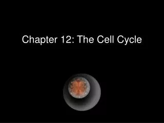
Reproduction – Sexual The Malaysian cycad Cycascircinalis. Left photo shows the "cone" of a female plant with modified leaves (sporophylls) bearing small ovules along their margins. Center photo shows a female plant with clusters of mature seeds atached to the sporophylls. Right photo shows the erect, pollen-bearing cone (strobilus) of a male plant. The individual scales (sprophylls) of the cone bear clusters of sproangia.
Reproduction – Sexual • Strictly dioecious plant • Male plants are rare • Male strobilus or cone borne singly at the apex of the trunk • Apical shoot apex utilized in the development of male cone, hence branching sympodial • Cone shortly stalked & large (up to 50cm length or more)
Reproduction – Sexual • Numerous micro-sporophylls spirally arranged around the central axis • Each micro-sporophyll is narrow below and broad above terminating into projection – the apoplysis • Microsporangia confined to abaxial (lower) surface • Usually present in sori – each with 2-6 sporangia • They contain a large number of haploid microspores (pollen grains)
Female Reproductive Structures • Female plant do not produce definite cones • A whorl of spirally arranged megasporophylls arise around the short apex • Each megasporophyll resembles the foliage leaf and approximately 10-23 cms. long • Lower petiolar part bears the naked ovules on the margins
Ovule Structure • Largest ovule (6cms.x4cms.) seen in C.circinalis • Ovules are orthotropous, sessile, ovoid or spherical in shape and unitegmic. • The thick integument is differentiated in three layers-outer and inner fleshy layers, middle stony. • The integument remains fused inside with nucellar tissue except at the position where it forms the micropylar opening. • Ovule is well supplied with vascular bundles.
Megasporangium • The megaspore develops in the nucellus by meiotic division and goes on to form female gametophyte tissue. • 2-3 archegonia are formed in this haploid tissue which is food laden. • Egg cell in the venter of archegonia, undergoes fertilization by the motile spermatozoid forming diploid zygote.
Pollination - Development of male gametophyte after pollination • The pollen grains are carried by wind (Anemophily) and caught by pollination drop secreted by ovule. Pollination is direct. • The pollination drop is dehydrated and the pollen grains are sucked into the pollen chamber. • Pollen grains take rest for some time in the pollen chamber.
Pollination - Development of male gametophyte after pollination • During the germination of pollen grain the exine is ruptured and the inner intine comes out in the form a tube like structure known as pollen tube. • At this time the generative cell divides and forms a larger, upper body cell and smaller, lower stalk cell. • The pollen tube acts as haustorium to absorb food materials from the nucellus besides as sperm carrier. • The body cell divides and forms two naked, top shaped, motile, multiciliatedantherozoids. The cilia are in 4 – 5 spirals. • The male gametes of Cycas are 180 – 210 μ in size and largest in the plant kingdom. • The pollen tube apex is ruptured and the male gametes are released into the archegonial chamber. • Presence of multiciliated male gametes is the fern character shown by Cycas male gametophyte
Young Sporophyte – Embryo • Embryo development is meroblastic. • Proembryo shows upper haustorial part, middle elongating suspensors and the basal meristematicembryonalregion.
Seed • A mature embryo is straight and has a short hypocotyl. • Embryonal axis has plumule at one end and radicle at the other. • Radicle is covered by coleorhiza. • Number of cotyledons maybe 2-3.. • Nucellusis completely absorbed in the seed. • Mature seed is large 2.5–5 cm wide and usually orange or red in colour.. • Germination is hypogeal type.