Download
1 / 63
650 likes | 897 Vues
Lecture 15 plasma membrane transport Active transport pp72-79. Active Processes. Whenever a cell uses the bond energy of ATP to move solutes across the membrane, the process is referred to as active.

E N D
Active Processes • Whenever a cell uses the bond energy of ATP to move solutes across the membrane, the process is referred to as active. • Substances moved actively across the plasma membrane are usually unable to pass in the necessary direction by passive transport processes. The substance may be too large to pass through the channels, incapable of dissolving in the lipid bilayer, or unable to move down its concentration gradient.
Membrane Transport: Active Processes • Two types of active processes: • Active transport • Vesicular transport • Both use ATP to move solutes across a living plasma membrane
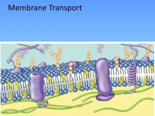
Active transport • like carrier-mediated facilitated diffusion, requires carrier proteins that combine specifically and reversibly with the transported substances. However, facilitated diffusion always honors concentration gradients because its driving force is kinetic energy. In contrast, the active transporters or solute pumps move solutes, most importantly ions (such as Na+, K+, and Ca2+), “uphill” against a concentration gradient. To do this work, cells must expend the energy ofATP.
Lipid-insoluble solutes (such as sugars or amino acids) (b) Carrier-mediated facilitated diffusion via a protein carrier specific for one chemical; binding of substrate causes shape change in transport protein Figure 3.7b
Active Transport • Requires carrier proteins (solute pumps) • Moves solutes against a concentration gradient • Types of active transport: • Primary active transport • Secondary active transport
Primary Active Transport • Energy from hydrolysis of ATP causes shape change in transport protein so that bound solutes (ions) are “pumped” across the membrane
Primary Active Transport • Sodium-potassium pump (Na+-K+ ATPase) • Located in all plasma membranes • Involved in primary and secondary active transport of nutrients and ions • Maintains electrochemical gradients essential for functions of muscle and nerve tissues
Extracellular fluid Na+ Na+-K+ pump Na+ bound K+ ATP-binding site Cytoplasm 1 Cytoplasmic Na+ binds to pump protein. P ATP K+ released ADP 6 2 K+ is released from the pump proteinand Na+ sites are ready to bind Na+ again.The cycle repeats. Binding of Na+ promotesphosphorylation of the protein by ATP. Na+ released K+ bound P Pi K+ 5 3 K+ binding triggers release of thephosphate. Pump protein returns to itsoriginal conformation. Phosphorylation causes the protein tochange shape, expelling Na+ to the outside. P 4 Extracellular K+ binds to pump protein. Figure 3.10
Extracellular fluid Na+ Na+-K+ pump K+ ATP-binding site Cytoplasm 1 Cytoplasmic Na+ binds to pump protein. Figure 3.10 step 1
Na+ bound P ATP ADP 2 Binding of Na+ promotesphosphorylation of the protein by ATP. Figure 3.10 step 2
Na+ released P 3 Phosphorylation causes the protein tochange shape, expelling Na+ to the outside. Figure 3.10 step 3
K+ P 4 Extracellular K+ binds to pump protein. Figure 3.10 step 4
K+ bound Pi 5 K+ binding triggers release of thephosphate. Pump protein returns to itsoriginal conformation. Figure 3.10 step 5
K+ released 6 K+ is released from the pump proteinand Na+ sites are ready to bind Na+ again.The cycle repeats. Figure 3.10 step 6
Extracellular fluid Na+ Na+-K+ pump Na+ bound K+ ATP-binding site Cytoplasm 1 Cytoplasmic Na+ binds to pump protein. P ATP K+ released ADP 6 2 K+ is released from the pump proteinand Na+ sites are ready to bind Na+ again.The cycle repeats. Binding of Na+ promotesphosphorylation of the protein by ATP. Na+ released K+ bound P Pi K+ 5 3 K+ binding triggers release of thephosphate. Pump protein returns to itsoriginal conformation. Phosphorylation causes the protein tochange shape, expelling Na+ to the outside. P 4 Extracellular K+ binds to pump protein. Figure 3.10
Secondary Active Transport • Depends on an ion gradient created by primary active transport • Energy stored in ionic gradients is used indirectly to drive transport of other solutes
Secondary Active Transport • Cotransport—always transports more than one substance at a time • Symport system: Two substances transported in same direction • Antiport system: Two substances transported in opposite directions
Symport • carries 2 or more solutes thru the membrane simultaneously in the SAME direction • Cotransport = process
Antiport • Carriers 2 or more solutes in the OPPOSITE directions • Counter transport = process
Extracellular fluid Glucose Na+-glucose symport transporter loading glucose from ECF Na+-glucose symport transporter releasing glucose into the cytoplasm Na+-K+ pump Cytoplasm 1 2 The ATP-driven Na+-K+ pump stores energy by creating a steep concentration gradient for Na+ entry into the cell. As Na+ diffuses back across the membrane through a membrane cotransporter protein, it drives glucose against its concentration gradient into the cell. (ECF = extracellular fluid) Figure 3.11
Extracellular fluid Na+-K+ pump Cytoplasm 1 The ATP-driven Na+-K+ pump stores energy by creating a steep concentration gradient for Na+ entry into the cell. Figure 3.11 step 1
Extracellular fluid Glucose Na+-glucose symport transporter loading glucose from ECF Na+-glucose symport transporter releasing glucose into the cytoplasm Na+-K+ pump Cytoplasm 1 2 The ATP-driven Na+-K+ pump stores energy by creating a steep concentration gradient for Na+ entry into the cell. As Na+ diffuses back across the membrane through a membrane cotransporter protein, it drives glucose against its concentration gradient into the cell. (ECF = extracellular fluid) Figure 3.11 step 2
Vesicular Transport • Transport of large particles, macromolecules, and fluids across plasma membranes • Requires cellular energy (e.g., ATP)
Vesicular Transport • Functions: • Exocytosis—transport out of cell • Endocytosis—transport into cell • Transcytosis—transport into, across, and then out of cell • Substance (vesicular) trafficking—transport from one area or organelle in cell to another
Endocytosis and Transcytosis • Involve formation of protein-coated vesicles • Often receptor mediated, therefore very selective
Types • Endocytosis – vesicular process that brings matter INTO the cell • Exocytosis – vesicular process that releases matter OUTSIDE the cell
Exocytosis Figure 3.12a
Vesicular Transport • Transcytosis – moving substances into, across, and then out of a cell • Vesicular trafficking – moving substances from one area in the cell to another • Phagocytosis – pseudopods engulf solids and bring them into the cell’s interior
Vesicular Transport • Fluid-phase endocytosis – the plasma membrane infolds, bringing extracellular fluid and solutes into the interior of the cell • Receptor-mediated endocytosis – clathrin-coated pits provide the main route for endocytosis and transcytosis • Non-clathrin-coated vesicles – caveolae that are platforms for a variety of signaling molecules
Coated pit ingestssubstance. 1 Extracellular fluid Plasmamembrane Protein coat(typicallyclathrin) Cytoplasm 2 Protein-coatedvesicledetaches. 3 Coat proteins detachand are recycled toplasma membrane. Transportvesicle Endosome Uncoatedendocytic vesicle 4 Uncoated vesicle fuseswith a sorting vesiclecalled an endosome. 5 Transportvesicle containingmembrane componentsmoves to the plasmamembrane for recycling. Lysosome 6 Fused vesicle may (a) fusewith lysosome for digestionof its contents, or (b) deliverits contents to the plasmamembrane on theopposite side of the cell(transcytosis). (b) (a) Figure 3.12
Coated pit ingestssubstance. 1 Extracellular fluid Plasmamembrane Protein coat(typicallyclathrin) Cytoplasm Figure 3.12 step 1
Coated pit ingestssubstance. 1 Extracellular fluid Plasmamembrane Protein coat(typicallyclathrin) Cytoplasm 2 Protein-coatedvesicledetaches. Figure 3.12 step 2
Coated pit ingestssubstance. 1 Extracellular fluid Plasmamembrane Protein coat(typicallyclathrin) Cytoplasm 2 Protein-coatedvesicledetaches. 3 Coat proteins detachand are recycled toplasma membrane. Figure 3.12 step 3
Coated pit ingestssubstance. 1 Extracellular fluid Plasmamembrane Protein coat(typicallyclathrin) Cytoplasm 2 Protein-coatedvesicledetaches. 3 Coat proteins detachand are recycled toplasma membrane. Endosome Uncoatedendocytic vesicle 4 Uncoated vesicle fuseswith a sorting vesiclecalled an endosome. Figure 3.12 step 4
Coated pit ingestssubstance. 1 Extracellular fluid Plasmamembrane Protein coat(typicallyclathrin) Cytoplasm 2 Protein-coatedvesicledetaches. 3 Coat proteins detachand are recycled toplasma membrane. Transportvesicle Endosome Uncoatedendocytic vesicle 4 Uncoated vesicle fuseswith a sorting vesiclecalled an endosome. 5 Transportvesicle containingmembrane componentsmoves to the plasmamembrane for recycling. Figure 3.12 step 5
Coated pit ingestssubstance. 1 Extracellular fluid Plasmamembrane Protein coat(typicallyclathrin) Cytoplasm 2 Protein-coatedvesicledetaches. 3 Coat proteins detachand are recycled toplasma membrane. Transportvesicle Endosome Uncoatedendocytic vesicle 4 Uncoated vesicle fuseswith a sorting vesiclecalled an endosome. 5 Transportvesicle containingmembrane componentsmoves to the plasmamembrane for recycling. Lysosome 6 Fused vesicle may (a) fusewith lysosome for digestionof its contents, or (b) deliverits contents to the plasmamembrane on theopposite side of the cell(transcytosis). (b) (a) Figure 3.12 step 6
Endocytosis • Phagocytosis—pseudopods engulf solids and bring them into cell’s interior • Macrophages and some white blood cells
(a) Phagocytosis The cell engulfs a large particle by forming pro- jecting pseudopods (“false feet”) around it and en- closing it within a membrane sac called a phagosome. The phagosome is combined with a lysosome. Undigested contents remain in the vesicle (now called a residual body) or are ejected by exocytosis. Vesicle may or may not be protein- coated but has receptors capable of binding to microorganisms or solid particles. Phagosome Figure 3.13a
Endocytosis • Fluid-phase endocytosis (pinocytosis)—plasma membrane infolds, bringing extracellular fluid and solutes into interior of the cell • Nutrient absorption in the small intestine
(b) Pinocytosis The cell “gulps” drops of extracellular fluid containing solutes into tiny vesicles. No receptors are used, so the process is nonspecific. Most vesicles are protein-coated. Vesicle Figure 3.13b
Endocytosis • Receptor-mediated endocytosis—clathrin-coated pits provide main route for endocytosis and transcytosis • Uptake of enzymes low-density lipoproteins, iron, and insulin
(c) Receptor-mediated endocytosis Extracellular substances bind to specific receptor proteins in regions of coated pits, enabling the cell to ingest and concentrate specific substances (ligands) in protein-coated vesicles. Ligands may simply be released inside the cell, or combined with a lysosome to digest contents. Receptors are recycled to the plasma membrane in vesicles. Vesicle Receptor recycled to plasma membrane Figure 3.13c
Exocytosis • Examples: • Hormone secretion • Neurotransmitter release • Mucus secretion • Ejection of wastes
Summary of Active Processes • Also see Table 3.2
Membrane Potential • Separation of oppositely charged particles (ions) across a membrane creates a membrane potential (potential energy measured as voltage) • Resting membrane potential (RMP): Voltage measured in resting state in all cells • Ranges from –50 to –100 mV in different cells • Results from diffusion and active transport of ions (mainly K+)