Soft Tissue Neck
Soft Tissue Neck. Presented to you by……. Angela Adolfs, Shannah Herron, Marissa Klein All information gathered at St. Joseph’s Hospital’s Radiology Department and from Dr. Bretzke . Equipment used: Siemens Somatom Definition Flash, Helical Scanner, 64 slice scanner.
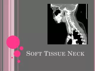

Soft Tissue Neck
E N D
Presentation Transcript
Presented to you by…….. • Angela Adolfs, Shannah Herron, Marissa Klein • All information gathered at St. Joseph’s Hospital’s Radiology Department and from Dr. Bretzke. • Equipment used: Siemens Somatom Definition Flash, Helical Scanner,64 slice scanner
General Clinical Indications Clinical Indications for ordering a STN include: • masses occurring in the neck • foreign bodies in the neck • laryngeal cancer • swollen lymph nodes • general inflammation or swelling • parathyroid adenoma • parotid gland mass
Description of Examination STN CT exam is a scan of the soft tissue of the neck. It scans from frontal sinuses to the aortic arch. The anatomy demonstrated includes: muscles, throat, tonsils, adenoids, airway, thyroid, and other glands. If contrast is to be used and an IV is not already present, an IV is started to by the technologist to administer contrast
Description of Examination • Once the IV is set, a quick scout is taken to determine patient position in table and the scan field of view is selected by the technologist. • When the patient is ready to be scanned, the patient table will slowly move through the gantry. • It is important that the patient remain still.
Description of Examination • The Patient is scanned after contrast is administered. • Once the scan is complete, all the necessary information can be gathered and reconstructed by the technologist.
Patient Preparation • If the scan does not require contrast, there is no patient preparation needed.
Patient Preparation If the patient is over 55, a diabetic, has a history of renal disease, or is being administered chemotherapy, then a creatinine needs to be checked before contrast is administered. Creatinine level along with age and other factors are used to calculate the GFR. If Contrast is to be administered, the patient should be NPO 2 hours prior to the exam.
Patient Preparation • Technologist discusses the patient’s history with patient, including: • any medications • any allergies that might cause a reaction to contrast • past medical history that would cause the images to have abnormal results • If the patient is on metformin, they need to be taken off for 2 days after the exam. • This is to ensure that the kidneys are not over worked. • Blood tests will determine if they can resume their metformin.
Patient Preparation Patient must remove all jewelry or metal on or near the head, neck and chest before the exam can be started. The patient will then lie down on the table with there head closest to the gantry. Their head will rest in the head rest provided. Once the patient is set, the technologist can move the patient table into position. To ensure that the right area will be scanned, the technologist will center the horizontal laser light at the hair line. The vertical laser light will be centered at the midaxillary line. Patient will slightly extend their chin to help project the mandible out of the way of the neck.
Contrast Media • Contrast is used to differentiate between blood vessels, vascular tumors and lymph nodes. • CTA or CT Angiography produces detailed images of blood vessels using contrast. An example of its use is to look for aneurisms. • CTV or CT Venography uses contrast to produce detailed images specifically of veins. An example of its use is to look for a thrombosis or clot.
Contraindications for Contrast • Allergy / Hypersensitivity to Iodine • Anuria • High Creatinine Level • GFR (Glomerular filtration rate) greater than: • >60 use normal amount of CM • 50-60 reduce dose, water intake 24 hrs • 40-50 reduce dose, hydrate 250-500 mL saline (pre/post) • 30-40 confirm for no contrast • Renal Disease or other conditions that compromise renal function (e.g. diabetes mellitus, multiple myeloma, sickle cell anemia, pheochromocytoma, lupus) • Congestive heart failure • Severe dehydration
Contrast Preparation • MEDRAD’s Stellant Injection System uses power injection to inject contrast media for enhanced visualization in CT diagnostic imaging procedures. • The amount of contrast and saline that needs to be administered can be adjusted based on the exam being done. • Contrast is warmed to decrease viscosity.
Contrast Media- Soft Tissue Neck CT • The most common contrast used for a STN is 100 mL of Omnipaque 350 with 100 mL of saline. • The contrast is injected at a rate of 2mL/sec.
Contrast Media- Soft Tissue Neck CT • Scan delay is set for 140 seconds • At 0 second, the technologist will flush the saline and start the 1st half of contrast -this will show glands or masses enhanced • At 90 seconds the 2nd half of contrast is administered • this will show enhanced vessels • the scan begins after 140 seconds
Contrast Media • Areas of the neck that are enhanced include: -Blood vessels, lymph nodes and any possible masses. -All tissues are slightly enhanced • Areas that remain unenhanced include: -Muscles • Pathology shown on an enhanced scan: -Abscesses, Abnormal gland tissue, Blood
Example of Contrast Enhanced STN showing masses • This Contrast-enhanced CT demonstrates masses
Patient Positioning Patient lies supine with a cushion under knees to support back Arms down at the patient’s sides Patient is velcroed onto the table with velcro straps to ensure their safety Once secure, the table is lifted and moved into the gantry to the proper alignment that is set by the technologist Patient will be directed to suspend breathing and swallowing
Scout Scan • A scout image is taken to determine patient position in table and the scan field of view is selected.
Scanning protocol • Mode: Helical Scanning Mode • Range: Frontal sinuses to Aortic arch or TEA to T1 • Thickness: 3.0 mm • SFOV: 200 (this is based off the size of the patient) • Exposure: 100kV, 187 Eff mAs • Scan time/Table Feed: -27.81 seconds/Craniocaudal table feed
Post processing • DFOV: 200 mm • Images are reconstructed using preset window width and levels. • Multiplanar reconstruction is used to produce Coronal and Sagittal views. A soft tissue window is applied to the Coronal and Sagittal views. • An Axial Lung window is also reconstructed.
Case #1PeritonsillarAbscess • 37 y/o male with a left side abscess that begins in the region of the left palantine tonsil.
Disease process and etiology • A Peritonsillar abscess is the most common infection of head and neck in adults. • An abscess is a collection of puss. • It is most commonly caused by the bacteria Streptococcus pyogeness.
Clinical Indications The patient presented with neck pain and difficulty breathing Other typical symptoms are: Fever Trismus(jaw spasms) Difficulty swallowing Ear pain on same side as abscess
RISK FACTORS • Risk factors- patients with chronic tonsillitis or multiple trials of oral antibiotic may be predisposed to get a peritonsillarabscess. • Other risk factors: • chronic dental infections • smoking • chronic lymphocytic leukemia • stones or calcium deposits in the tonsils • Most common in 20-40 year olds. • Both genders are affected equally.
CT findings • A large abscess is identifiable by area of increased radiolucency. • The abscess begins at the left palentinetonsil and extends inferiorly to the level of the hyoid bone. • The abscess continues down into the left laryngeal vestibule. • At the glottis, the airway is not identified.
Surgical / medical treatment options • Airway risks were discussed with patient and the abscess was drained outside of CT. • Needle aspiration was performed to drain the abscess. • After aspiration, antibiotics and other medications may be given. • In advanced cases incision and drainage or tonsillectomy may be required.
Other diagnostic tests • Physical exam to check for trismus (lockjaw) from swelling and spasms of pterygoidmuscle. • Ultrasound • X-ray • Fluids that are aspirated will be sent to lab for a gram stain and cultures in order to identify the microorganism.
Prognosis • Individualized treatment is most successful. • Follow up CT is recommended. • If aspiration is successful, no further surgical management is required. • If there is a reoccurrence of tonsillitis, it is recommended that within 3-6 months the patient should schedule a tonsillectomy.
Post Aspiration Follow Up • After aspiration and a second CT, the findings show that the infection is still extensive at the abscessed area and is moderately smaller than on previous CT scan, this is due to the abscess drainage. • Follow up care is needed to monitor the abscess.
Hematoma - Case #2 • Hematoma is identified in the hypopharynx • It is located from the angle of the mandible to the tracheostomy
Patient Clinical Indications and Symptoms 58 y/o male presenting with pain in neck Typical symptoms of hematomas include: swelling redness pain Symptoms also differ depending on the site of hematoma and the structures near it
Cause • Hematomas can be caused by trauma or can be spontaneous. • This hematoma followed a total laryngectomy and hypopharyngeal graft in the operating room.
CT Findings • A 54.4 x 47.2 mm hematoma was found beginning at the angle of the mandible to above the tracheostomy • Extravasation of contrast is noted in the hematoma suggesting active bleeding • Extravasation is adjacent to the right external carotid branches which is the source of the bleeding
medical treatment options • Some hematomas like those of the skin and soft tissues may be treated with R.I.C.E. • Rest stands for: • Rest • Ice • Compression • Elevation • Treatment for a hematoma involving organs or some vascular structures often require immediate surgery to treat the hematoma and insure there is not permanent damage done. • This hematoma would have been treated with R.I.C.E. since it is above the airway.
Other diagnostic tests to confirm diagnosis • Physical exam is usually performed first • Ultrasound • X-ray
Prognosis • Due to the location of the hematoma, there was no need to surgically address the hematoma. Had the Hematoma occurred below the tracheostomy, the surgery staff would have address the hematoma right way. • R.I.C.E. would be followed for a treatment plan.
Radiologist conclusions • Dr. Bretzke stated that the hematoma was rather large. • It starts on the right side and at its largest, crosses midline. • There is a fluid level within the hematoma. • This is due to the cells and serum separating within the hematoma.
Conclusion CT is the modality of choice for soft tissue neck pathology due to its quick acquisition speed and spatial resolution. STN CT’s allow for the identification of anatomy and pathology in the neck including the visualization of vessels, muscles, glands, lymph nodes, masses and foreign bodies. STN CT’s aid in the treatment planning and in the monitoring of the response of the pathology in the neck.
Anatomy Quiz Label 1-10 1. 2. 3. 4. 5. 6. 7. 8. 9. 10.
Resources • www.aafp.org/afp/2002/0101/p93.html • CT techs at St. Joseph’s Hospital: Beth, Laurel and Jan • Notes from Class • http://www.two-views.com/CT_scan/Neck.html • http://www.medicinenet.com/hematoma/article.htm • http://bioweb.uwlax.edu/bio203/s2007/falk_pete/