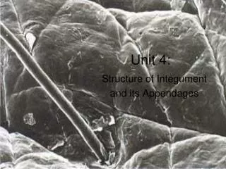
Unit 4:
Unit 4:. Structure of Integument and its Appendages. Regions of Integument (skin). Epidermis – outermost region Keratinized stratified squamous epithelium Non-vascular 4 cell types 4 - 5 layers. 4 Epidermal Cell Types. Keratinocytes – make keratin fibrous protein Protective

Unit 4:
E N D
Presentation Transcript
Unit 4: Structure of Integument and its Appendages
Regions of Integument (skin) • Epidermis – outermost region • Keratinized stratified squamous epithelium • Non-vascular • 4 cell types • 4 - 5 layers
4 Epidermal Cell Types • Keratinocytes – make keratin fibrous protein • Protective • Hardens and waterproofs skin. • Cells connected by desmosomes: • prevent tearing and cell separation from mechanical stress • Arise from hightly mitotic stratum basale • Cells dead at free surface
4 Epidermal Cell Types Keratinocytes – make keratin fibrous protein • Langerhans’ Cells – star shaped, epidermal dendritic phagocytic cells • activate the immune system • ingest foreign material
4 Epidermal Cell Types Keratinocytes – make keratin fibrous protein Langerhans’ Cells – star shaped, epidermal dendritic phagocytic cells • Merkel Cells – half-sun touch receptors • associated w/ sensory nerve endings
4 Epidermal Cell Types Keratinocytes – make keratin fibrous protein Langerhans’ Cells – star shaped, epidermal dendritic phagocytic cells • Merkel Cells – half-sun touch receptors • associated w/ sensory nerve endings melanin • Melanocytes – makesbrown pigment melanin • shields keratinocyte DNA from UV damage
Layers of the Epidermis Stratum corneum Stratum lucidum (absent in thin skin) Stratum granulosum Stratum Spinosum Stratum basale
Stratum Basale (Basal Layer) AKA stratum germinativum Deepest epidermal layer, attached to the dermis Single row of the youngest keratinocytes Rapidly mitotic, making new cells daily Melanocytes and Merkel cells found here Stratum basale dermis
Stratum Spinosum (Prickly Layer) Cells filled with filaments connected to desmosomes. (gives prickly look) Melanin granules filling cells in response to UV or genetics Langerhans’ cells found here Stratum Spinosum Stratum basale dermis
Stratum Granulosum (Granular) 3-5 cell layers Keratinocytes change, flatten, lose nuclei Keratin granules accumulate in the cells of this layer Lamellated granules release extracellular glycolipids in intercellular space that waterproof skin Too far from nutrient rich dermal blood, cells begin to die Stratum granulosum Stratum Spinosum Stratum basale dermis
Stratum Lucidum (Clear Layer) Transparent band of flat, dead keratinocytes Only in thick skin Sole of feet, palms, calluses Reduces friction between the granulosum (inferior) and the corneum (superior) Stratum lucidum would be here, if present Stratum granulosum Stratum Spinosum Stratum basale dermis
Stratum Corneum (Horny Layer) 20-30 layers of DEAD keratinized cells; ¾ of epidermal thickness Functions include: Waterproofing (due to glycolipids) Protection from: Abrasion Penetration biological, chemical, and physical assaults Stratum corneum Stratum lucidum would be here, if present Stratum granulosum Stratum Spinosum Stratum basale dermis
Stratum Corneum (Horny Layer) Stratum Corneum Can Little Stratum Lucidum Girls Stratum Granulosum Stratum Spinosum Smell Bad? Stratum Basale dermis
Let’s take a break from lecture to draw the difference between thick and thin skin. • Use appropriate drawing and coloring methods. • Horizontal labeling with leader lines http://www.lab.anhb.uwa.edu.au/mb140/corepages/integumentary/integum.htm#labepidermis
Regions of Integument (skin) • Epidermis – outermost region • Keratinized stratified squamous epithelium • Non-vascular • 4 cell types • 4 - 5 layers • Dermis – middle region • Vascularized • 80% dense irregular connective tissue • 20% areolar connective tissue
Overview of the Dermis Cell types: fibroblasts, phagocytes, mast cells and white blood cells 2 layers papillary (upper) and reticular (lower) Rich with nerves, blood and lymph vessels Most hair follicles, oil and sweat glands derived here
Papillary Layer of Dermis • Dermal papillae with: • capillary loops (thin blood vessels) • Meissner’s corpuscles (touch), • and free nerve endings (pain) • Areolar connective tissue with collagen and elastic fibers • Superior surface with dermal papillae: peg-like projections (reason for fingerprints)
Papillary Layer of Dermis • Krause’s end bulb: (cold) • Ruffini end organs: (heat and sustained pressure) • In Reticular Layer: • Pacinian corpuscle: (touch, deep vibrations, transient pressure)
Reticular Layer of the Dermis 80% of the thickness of the dermis (dense –irregular CT) Collagen fibers: add strength and resiliency Binds water, keeping skin hydrated Elastin fibers: stretch-recoil properties Rich in blood vessels: dilate or constrict in response to emotions or temperature changes
Name the epidermal and dermal layers (review) • 5. Stratum Corneum (Epidermis) • 4. Stratum Lucidum (Epidermis) • 3. Stratum Granulosum (Epidermis) • 2. Stratum Spinosum (Epidermis) • Stratum Basale (Epidermis) • 6. Papillary Layer (Dermis) • 7. Reticular Layer (Dermis)
Regions of Integument (skin) • Epidermis – outermost region • Keratinized stratified squamous epithelium • Non-vascular • 4 cell types • 4 - 5 layers • Dermis – middle region • Vascularized • 80% dense irregular connective tissue • 20% areolar connective tissue • Hypodermis (superficial fascia) • deepest region • Mostly adipose (fat storage), some areolar • Vascularized
Hypodermis(superficial or subcutaneous fascia) Composed mostly of adipose and some areolar connective tissue Adipose cells swell and thicken with fatty droplets during weight gain Connects skin to underlying muscle Absorbs shock Insulates
Skin Color Three pigments contribute to skin color Melanin: yellow to reddish-brown to black only pigment made in skin by melanocytes and passed onto keratinocytes Freckles and pigmented moles – result from local accumulations of melanin Carotene: yellow to orange pigment Pigment incorporated into skin due to diet Accumulates in stratum corneum and in adipose Hemoglobin: reddish pigment, gives pink hue to skin Due to oxygenation of red blood cells
Skin “Appendages” Epidermal Derivatives include: hair Sebaceous Oil Glands hair follicles 6 Sudoriferous Sweat Glands
Sudoriferous Sweat Glands(2 types: Eccrine and Apocrine) Eccrine glands Covers entire body (3 million p/person) Most abundant on palms, soles of the feet, and forehead Coiled in dermis Duct opens on skin’s surface (pore) “sweat” = hypotonic blood filtrate released by exocytosis: 99% water, salts, antibodies, anti-biotic proteins, and N-wastes, vitamin C Evaporation of sweat cools the body
Sudoriferous Sweat Glands(2 types: Eccrine and Apocrine) Apocrine glands Only 2000 p/person Found in axillary and anogenital areas with pheromone secretions Ducts empty into hair follicles Odorless initially. Secretions contains lipids and protein that bacteria feed on. Decomposition of secretions by bacteria produce “body odor”
Sweat glands modified Ceruminous glands – modified apocrine glands in external ear canal that secrete cerumen (ear wax) Mammary glands – specialized sweat glands that secrete milk
Sebaceous “Oil” Glands Simple branched alveolar glands Holocrine:glandular cells rupture to release secretions Sebum Secretions: Oils + ruptured cell fragments moisturize hair and skin Slows water-loss bactericidal Released onto hair within follicle then flows onto skin surface. Acne due to blockage of hair follicle w/ infected sebum
Video Summary You tube: What is skin? The Layers of Human Skin
Hair (Pili) • Strands of dead, hard-keratinized cells made by follicles • Softer keratin in epidermal cells • Shaft projects from skin; Root embeded within dermis and hypodermis • 3 concentric layers: • - Medulla: absent in fine hair • - Cortex: gives hair color • - Cuticle: overlapping keratin • Split ends: cuticle worn away, exposing cortex
Structure of Hair Follicle Follicle created by in-vagination of epidermal surface (epithelial root sheath) into dermis and hypodermis to create a “bag” or “sac” that builds hair Basement membrane medulla
Structure of Hair Follicle Deep end of follicle: expanded forming a hair bulb Hair papilla supplies nutrients to hair (via capillaries) and signals growth Melanocytes on superior surface of papilla pigments hair by creating melanin Hair papilla created from in-folding of dermal tissue into hair bulb
Structure of Hair Follicle Arrector pili muscle – attached to hair follicle and skin. When contracted, holds hair erect • Root hair plexus wraps around each hair bulb • Bending hair stimulates these endings, hence our hairs act as sensitive touch receptors
Hair Shape Internal shape of shaft and follicle determines hair shape Round shaft: straight hair Oval shaft: wavy hair Flat or ribbon like shaft: kinky, curly hair ** One head can have many shaft shapes resulting in interesting hair textures.
Hair Types Vellus – pale, fine body hair found in children and the adult female (immature) Terminal – coarse, long hair of eyebrows, scalp, axillary, and pubic regions Hair growth influenced by: Nutrition Blood flow: reduced blood flow hair loss Ex. Brick layer shoulders: increased blood flow to area because of carrying heavy objects results in hair growth
Hair Growth Cycles • Hair has a life cycle: • Period of Active Growth (AG) • Regressive Phase: hair bulb shrivels and matrix dies • Resting Phase • Cycle repeats: Older hair falls out, replaced by new hair • Length of AG period determine length of hair • Ex: • Scalp: AG of 6-10 years • Brows AG: of 3 – 4 months • Balding or thinning hair: short AG
Hair Thinning and Baldness Alopecia – hair thinning in both sexes Rate of hair shed > Rate of hair growth Hirsutism: excessive hair growth in women. Caused by excessive sex hormones usually from an ovarian tumor. True, or frank, baldness Genetic Sex-influenced condition Male pattern baldness – caused by follicular response to DHT (Dihydrotestosterone) Growth cycle is so short that hairs never emerge from follicles before shedding Sex linked trait – carried on X chromosome, inherited from mother
Hair Function Functions of hair include: maintaining warmth Alerting the body to insects on skin Guarding the scalp against trauma, heat loss, and sunlight Eyelashes and nose hairs act as barriers against foreign substances
Hair Distribution Hair is distributed over the entire skin surface except: Palms, soles, and lips Nipples and portions of external genitalia
Structure of a Nail Scale-like epidermal modification on the distal, dorsal surface of fingers and toes w/ hard keratin Figure 5.6
Back to Regions of Skin 20% Areolar Connective Tissue 80% Dense Irregular Connective Tissue • Collagen fibers organized in irregular patterns • Strong and flexible
Back to Regions of Skin Adipose Tissue of Hypodermis Mostly fat droplets Dense irregular connective tissue of reticular layer of dermis Adipose tissue of hypodermis