Spirochete Diseases
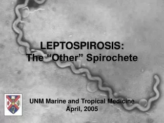
Spirochete Diseases. Terry Kotrla, MS, MT(ASCP)BB Fall 2005. Introduction to Spirochetes. Long, slender, helically tightly coiled bacteria Gram-negative Aerobic, microaerophilic or anaerobic . Corkscrew motility Can be free living or parasitic

Spirochete Diseases
E N D
Presentation Transcript
Spirochete Diseases Terry Kotrla, MS, MT(ASCP)BB Fall 2005
Introduction to Spirochetes • Long, slender, helically tightly coiled bacteria • Gram-negative • Aerobic, microaerophilic or anaerobic . • Corkscrew motility • Can be free living or parasitic • Best-known are those which cause disease: Syphilis and Lyme’s disease
Morphology • Have axial filaments, which are otherwise similar to bacterial flagella • Filaments enable movement of bacterium by rotating in place
Spirochete Diseases • Localized skin infection disseminates to other organs. • Latent stage, no signs/symptoms apparent. • Cardiac and neurological involvement in untreated cases.
Serological Testing • Important in diagnosis • Isolation of organism very difficult • Clinical symptoms not always apparent.
Syphilis • Most commonly acquired spirochete disease in the U.S. • Complex sexually transmitted disease that has a highly variable clinical course. • In 2004, syphilis cases reported to CDC increased to 7,980 from 7,177 in 2003, an increase of 11.2%.
Primary and secondary syphilis - Rates by state: United States and outlying areas, 2004
Primary and secondary syphilis - Rates by county: United States, 2004
Characteristic of the Organism • Causative agent is Treponema pallidum • Member of the family Spirochaetaceae. • No natural reservoir in the environment, requires living host. • Organism cannot be cultured from clinical specimens
Morphology • Spiral shaped and motile due to periplasmic flagella. • Variable length.
Other Treponemes • Three other pathogens in the group: Treponema which are morphologically and antigenically similar to T. Pallidum • Differences are in: • characteristics of lesions, • Amount of systemic involvement and • course of the disease.
T. pertenue • Found in tropics, causes disease Yaws. • Non-venereal transmission, transmitted by direct contact. • Disease of bone and skin, rarely viscera • Persistent lesions, wart-like, occur primarily in children, causes and ulcerative necrosis, scar formation, disfiguring. • Untreated disease not as severe as syphilis, but lesions are more persistent. • Treat with penicillin • Serologic syphilis test will be reactive.
T. pertenue • Occurs mainly in equatorial regions and can be found in South America, Central America, the Caribbean, Africa, and Southeast Asia. • It is associated with high humidity and rainfall. • Fifty years ago, the WHO recognized that endemic treponematoses—yaws in particular—were a major cause of disfigurement and disability and a significant economic burden in poor countries.
Infection with Treponema pallidum pertenue. Notice the deformed tibiae, the so-called sabre tibiae.
T. endemicum • Causes non-venereal syphilis known as bejel or endemic syphilis • Typically spread among children, most commonly in the Middle East and the southern Sahara desert regions. • Bejel is completely curable with penicillin. • Serologic syphilis test will be reactive.
T. endemicum • Bejel affects the skin, bones, and mucous membranes of the mouth. • Transmission is by direct contact, with broken skin or contaminated hands, or indirectly by sharing drinking vessels and eating utensils. • Symptoms begin with a slimy patch on the inside of the mouth followed by blisters on the trunk, arms, and legs. • Bone infection develops later, mainly in the legs. • Also in later stages, soft, gummy lumps may appear in the nose and on the roof of the mouth (soft palate).
T. caroteum • Pinta (T carateum) occurs in Central and South America and the Caribbean. • More common in young adults. • Non-venereal, direct contact, disease of skin. • Lesion is initially a scaly patch, becomes red-blue, later becomedepigmented and atrophy. • Treat with penicillin • Serologic syphilis test will be reactive.
Pinta • Depigmented skin lesions.
T. cuniculi • Not pathogenic for humans, • Causes rabbit syphilis.
Mode of Transmission • Organism is very fragile, destroyed rapidly by heat, cold and drying. • Sexual transmission most common, occurs when abraded skin or mucous membranescome in contact with open lesion. • Can be transmitted to fetus. • Rare transmission from needle stick and blood transfusion.
Stages of Disease • Primary • Secondary • Latent • Tertiary • Congenital Syphilis
Primary Syphilis • Organism enters directly through skin or through mucosal tissue. • Carried by blood throughout the body. • Organisms remaining at the site begin to multiply.
Primary Syphilis - Chancre • Variable incubation period of 10 days to several months, a primary lesion, chancre, forms at the entrance site. • Chancre begins as a small, usually singular nodule; as it enlarges, the overlying epithelial tissues begins to necrose, resulting in a relatively painless ulcer. • Unlike other bacterial infections, there is no formation of pus unless a secondary bacterial infection sets in.
Primary - Chancre • Chancre is most frequently seen on the external genitalia • In women the lesions may form in the vagina or on the cervix. • In men it may be inside the urethra, resulting in a serous discharge. • The lesion heals spontaneously after 1-5 weeks. • Swab of chancre smeared on slide, examined under dark-field microscope, spirochetes will be present. • Thirty percent become serologically positive one week after appearance of chancre, 90% positive after three weeks.
Secondary Syphilis • Occurs 6-8 weeks after initial chancre, becomes systemic, patient highly infectious. • Characterized by localized or diffuse mucocutaneous lesions, often with generalized lymphadenopathy. • Primary chancre may still be present. • Secondary lesions subside in about 2-6 weeks. • Serology tests nearly 100% positive.
Secondary Syphilis • A widespread eruption resembling psoriasis or pityriasis rosea which prominently involves the hands should always include the differential diagnosis of secondary syphilis.
Secondary Syphilis • Secondary syphilis lesions on back
Latent Syphilis • Stage of infection in which organisms persist in the body of the infected person without causing symptoms or signs (asymptomatic). • This stage may last for years. • One-third of untreated latent stage individuals develop signs of tertiary syphilis. • After four years it is rarely communicable sexually but can be passed from mother to fetus.
Latent Syphilis • This stage may be further subdivided. • Early latent, initial infection occurred within previous 12 months. • Late latent, initial infection occurred greater than 12 months. • Latent of unknown duration, date of initial infection cannot be established as having occurred in the previous year.
Tertiary Syphilis • Divided into three manifestations: • Gummatous syphilis • Cardiovascular syphilis • Neurosyphilis
Tertiary Syphilis - Gummatous • Gummas are localized areas of granulomatous inflammation found on bones, skin and subcutaneous tissue. • Cutaneous gummas may be single or multiple, generally asymmetric and grouped together. • Visceral lesions often cause local destruction of the affected organ. • Contain lymphocytes, plasma cells and perivascular inflammation.
Tertiary - Cardiovascular • This condition appears 20 or more years post-infection. • Usually involves the aorta. • Invading treponemes cause scarring of the tunica media. • Over many years, the inflammatory scarring weakens the aortic wall, leading to aneurysm formation, which causes incompetence of the aortic valve and narrowing of the coronary ostia.
Tertiary - Cardiovascular • Antibiotic treatment cures the syphilis infection and stops the progress of cardiovascular syphilis. • The damage that has already occurred may not be reversed.
Neurosyphilis • Caused by invasion of organisms into the CNS. • Manifests as an insidious but progressive loss of mental and physical functions and is accompanied by mood alterations. • General paresis of the insane: • forgetful, • personality change, • psychiatric symptoms. • Onset usually 10-20 years after primary infection. • Treatment may not improve symptoms.
Neurosyphilis • Neurological complications at this stage include generalized paresis of the insane which results in personality changes, changes in emotional affect, hyperactive reflexes. • Tabes dorsalis, degeneration of lower spinal cord, general paresis and chronic progressive dementia often results in a characteristic shuffling gait. • Can only be diagnosed serologically by VDRL.
Neurosyphilis • Cerebral atrophy, most prominent in frontal lobes seen in general paresis.