
BLOOD
BLOOD. Blood. Transports substances Maintains homeostasis Type of CT composed of cells w/in a noncellular matrix Hematophobia = fear of blood. Blood and Blood Cells. 2 components 1. cells ( rbc , wbc , platelets) 45%

BLOOD
E N D
Presentation Transcript
Blood • Transports substances • Maintains homeostasis • Type of CT composed of cells w/in a noncellular matrix • Hematophobia= fear of blood
Blood and Blood Cells • 2 components 1. cells (rbc, wbc, platelets) 45% 2. Plasma (water, amino acids, proteins, carbohydrates, lipids, vitamins, hormones, electrolytes, cellular waste) 55% Hematocrit- volume of blood cells in a sample, should be 45%. The remaining fluid is plasma 55%.
3 types of blood cells • Red blood cells- erythrocytes • White blood cells- leukocytes • Platelets- therombocytes
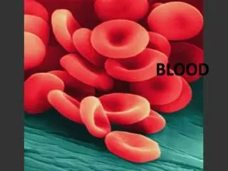
Red Blood Cells • Biconcave shape • 5 mil/ cubic millimeter • Lacking nuceli • Do not divide • Formed in bone marrow= Hemotopoeisis • Live 120 days phagocytized by liver and spleen
RBC Function • Transports O₂ throughout body, picks up CO₂ • Hemoglobin= molecule which combines with O₂ to transport it
Oxygen Levels • Oxyhemoglobin- oxygen rich- bright red • Deoxyhemoglobin-not carrying O₂- bluish red
Elements Critical to RBC Production • Folic Acid • Vitamin B12 • Iron- needed to synthesize hemoglobin • Anemia= too few RBC • Erythropoietin secreted by kidneys stimulates RBC formation
White Blood Cells • Function- defend the body against disease- causing agents • Granulocytes- granular cytoplasm • Agranulocytes- lacking granular cytoplasm
Granulocytes- 3 types • Neutrophils- very active in phagocytosis of bacteria and are present in large amount in the pus of wounds • 60% WBC- most common
2. Eosinophils- attack parasites • Control allergic reaction • 2 % WBC
3. Basophiles- produce Heparin(prevents blood clots) • Produces Histamine(causes inflammatory reaction) • 1% WBC
Agranulocytes- 2 1. Monocytes- precursors of macrophates • Phagocytosis- bacteria, debris, other cells • 6% WBC
2. Lymphocytes- main constituents of the immune system which is a defense against the attack of pathogenic microorganisms such as viruses, bacteria, fungi, and protista • Yield antibodies/ arrange them on their membrane • 30% WBC
Platelets (thrombocytes) • Help initiate formation of blood clots, close breaks in damaged vessels • Arise from cell in bone marrow called megakaryocytes • These cells fragment and release small sections (platelets) of cytoplasm into circulation • Less that ½ the size of a red blood vessel
HOMEWORK • Pg 530 1-3 • Pg 533 1-3 • Pg 534 1-2 • Pg 537 1 • Pg 539 2-4
Blood Plasma • Liquid portion of blood • 92% water • Transport nutrients, gases , vitamins, maintain fluid and electrolyte balance, and pH
Plasma Proteins 1. Albumins- made in liver , maintain osmotic pressure and blood volume(blood pressure) 2. Globulins- 3 groups: alpha, beta, gamma • Alpha& Beta- from liver, transport lipids and fat soluble vitamins • Gamma- from lymphatic tissues, antibodies for immunity 3. Fibrinogen- from liver, largest molecules of plasma proteins- important for blood clotting. Major event in blood clotting is the change of fibrogen into fibrin.
Hemostasis • Process of stopping bleeding • Coagulation causes the formation of blood clots • 3 key events
1. Blood vessel spasm- damaged vessels stimulate muscle tissue in wall of blood vessels to contract. Slows or stops blood flow, lasts several minutes. Platelets release serotonin, a vasoconstrictor which maintains the muscle spasm longer
Platelet plug formation- platelets stick to surfaces of damaged blood vessels and to each other to form a plug
3. Blood coagulation- most effective, forms a blood clot(hemotoma) . Injury causes an increase in the release of coagulants. Main event- conversion of fibrinogen into long protein threads called fibrin.
Tissue damage= production of prothrombin activator • Prothrombin- converted to thrombin • Thrombin acts as a enzyme to cause change of fibrinogen to fibrin, which trap platelets and blood cells to form a hemotoma • Thrombus= blood clot abnormally forming in a vessel • Embolus= clot moves and becomes lodged in another place
Coagulation- thickening of blood to form a clot • http://www.dnatube.com/video/2680/Hemostasis
Blood Diseases • Anemia- iron deficiency • Sickle Cell Anemia- genetic disorder • Sickle shaped blood cells • Pain, lethargy, organ failure, stroke
Leukemia- type of cancer • Overproduction of wbc- take place of rbc • treatable with bone marrow transplants, chemotherapy, radiation
Infectious Mononuclosis- • Mono- • viral infection
Blood Poisoning- Septicemia • infection enters blood stream- can be deadly • treated with antibiotics • Thrombocytopenia- low production of platelets • Bleeding and bruising
Hemophila- genetic disorder • Failure of blood to clot • Treated with blood transfusions that include clotting agents
Hemophilia is carried on the X chromosome • Females XHXH normal • XHXh carrier • XhXh hemophiliac • Males X HY normal • X h Y hemophiliac
Systemic Circulations- delivers blood to all body cells and carries away waste • Pulmonary circulation- eliminates CO₂ and oxygenates blood (lung pathway)
Structure of The Heart • Heart Size – about 14 cm x 9 cm (the size of a fist). • Located in the mediastinum • The distal end of the heart is called the apex.
Coverings of Heart • Pericardium- encloses the heart (like a bag) • Visceral- inner • Parietal- (outer, attached to diaphragm, sternum and vertebrae) • Pericardial cavity- contains fluid for the heart to float in, reducing friction
Wall of Heart • Epicardium– outer layer, reduces friction • Myocardium – middle layer, mostly cardiac muscle • Endocardium – thin inner lining, within chambers of the heart
Heart Chambers and Valves • Your heart is a double pump. Circulation is a double circuit: • Pulmonary (lungs only) • Systemic (rest of the body)
Heart has 4 chambers: • 2 Atria – thin upper chambers that receive blood returning to the heart through veins.. Right and Left Atrium • 2 Ventricles – thick, muscular lower chambers. Receive blood from the atria above them. Force (pump) blood out of the heart through arteries. Right and left ventricle. • Septum – separates the right and left sides of the heart
Valves of the Heart • – allow one-way flow of blood. 4 total • 2 Atrioventricular Valves (AV) • bicuspid valve or mitral valve- Between left atrium and ventricle • tricuspid valve- Between right atrium and ventricle • 2 semilunar Valves • Aortic Semilunar – or just aortic valve. Between the left ventricle and the aorta • Pulmonary Semilunar, or just pulmonary valve. Between the left ventricle and the aorta
Arteries/Veins • Superior and Inferior Vena Cava- lead to right atrium carrying deoxygenated blood from all parts of body. • Pulmonary Trunk- divides into left/right pulmonary arteries • Pulmonary Arteries- carry deoxygenated blood to lungs • Pulmonary Veins- bring oxygenated blood from lungs to left atrium • Aorta- large artery carrying oxygenated blood to body from left ventricle
Path of Blood through Heart • Deoxygenated blood enters right atrium through the vena cava • Deoxygenated Blood moves into the right ventricle • Deoxygenated Blood goes out the pulmonary arteries and heads to the lungs • Oxygenated Blood returns from lungs and enters the left atrium • Oxygenated Blood moves into the left ventricle • Oxygenated blood moves out of the left ventricle through the aorta and to the body
Superior vena cava Inferior Vena Cava
http://www.mydr.com.au/heart-stroke/animation-how-your-heart-pumpshttp://www.mydr.com.au/heart-stroke/animation-how-your-heart-pumps • http://www.nhlbi.nih.gov/health/dci/Diseases/hhw/hhw_pumping.html • http://www.wisc-online.com/objects/ViewObject.aspx?ID=AP12504
Skeleton of the Heart- dense connective tissue holding the heart and valves in place


