Download
1 / 1
10 likes | 138 Vues
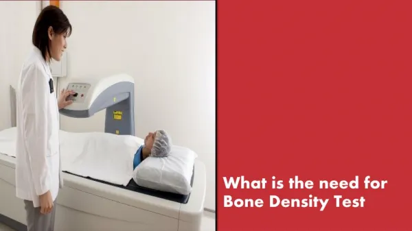
Investigating the relationship between density and magnetic susceptibility in bone samples to aid medical diagnostics. Results show minimal density changes post-immersion, suggesting in-vitro measurements approximate in-vivo density.

E N D
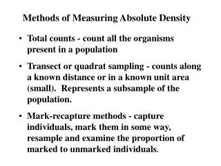
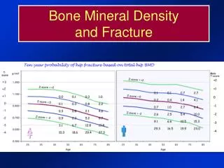
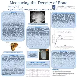
Kyle Strodtbeck Advisor: David Farrell • Department of Physics, CWRU, 10900 Euclid Ave., Cleveland, Ohio 44106 ABSTRACT Density and magnetic susceptibility are fundamental properties of condensed matter and some recent work [1] suggests that there may be a direct relationship between them in the case of biological materials. Magnetic susceptibility may therefore provide a useful tool to image the density of the human body, which could be of considerable value in medical diagnostics. However, probing this relationship quantitatively demands an accurate measurement of the in-vitro density of biological samples. Because of their porous structure, inhomogeneity, and loss of fluid after they are removed from the living organism, this is a challenging task. The present investigation is focused on bone, where the problems mentioned are all present but less severe than in soft tissue. Bone samples were collected for several different species and a specially designed container was utilized to probe their density when as a function of their time of immersion in water. We have discovered that very significant changes in both volume and mass can occur for (dry) bone following immersion but that these changes are correlated in a manner that leads to very small changes in density. Our results encourage the view that in-vitro density measurements of the sort described here provides a good approximation to the true in-vivo density of bone in living organisms. RESULTS AND DISCUSSION (continued) The results tabulated above divide naturally into two groups, namely, bones with relatively large (>15%) changes in density (mahi and groupie) and those with fairly small changes (<3%). Although we made no experiments on human bone, it is probable that the human case falls into the latter category, so we performed detailed time dependent studies of the mass and volume for three of the second group over shorter time periods, with the results graphed below. It is clear from these results that the mass and volume changes are rapid and substantial, and that the small density changes simply results from the fact that the mass and volume changes are closely correlated. Source: Uniformed Services University METHODS The traditional “Archimedes” approach to density measurement, in which the sample is immersed in water, was adopted for our measurements. A container was constructed that allow samples with relatively large volumes (on the order of 50 cm3) to be measured. Preliminary measurements were made to explore the difference between what we term the “dry” and “wet” state of our samples. In the former, samples were dried for 50 seconds at medium power in a household microwave oven. Their mass was measured, together with the volume of water displaced immediately following submersion. In the other extreme (“wet samples”) the density was measured after 24 hours of submerging the dry bone in water. These initial measurements were followed up with detailed time-dependent studies of the mass and volume for three of these samples over shorter time periods, using the same Archimedian technique. Measuring the Density of Bone CONCLUSION Relatively large changes in mass and volume occur following submersion of dry bone in water, but in most cases these changes essentially cancel each other out as far as the density is concerned. In fact, with the exception of bones obtained from the groupie and mahi fish, the overall change in density on immersion in water is just one or two percent. Physically, it appears that water does not flow into empty spaces in the structure, which would leave the volume unchanged, but simply expands the structure by an amount proportional to the volume of water absorbed. Independent of their detailed physical interpretation, our results encourage the view than an in-vitro density measurement of the sort described here provides a reliable approximation for the true in-vivo density of bone in a variety of living organisms. FIGURE ONE Density container with small, removable leak-tight top and black reference line Source: US National Cancer Institute INTRODUCTION Bones form part of the endoskeleton of vertebrates and are among the most diverse organs in the human body. Due to different hormone ratios, collagen disproportion, water content, and other physiological variability, bone density is expected, and found in previous studies [2] to vary significantly between different species. A further experimental difficulty attaches to in-vitro density measurements which are not present for the type of samples normally studied in condensed matter physics, namely, the loss of fluid attendant upon separating the bone from its parent organism. Recent work [1] has suggested that a direct relationship exists between the magnetic susceptibility and density of soft biological materials. An extended set of susceptibility and density measurements on bone samples from different species is needed to test whether there is a simple relationship between density and susceptibility for bone. RESULTS AND DISCUSSION Measurements for density were taken in both dry and wet states, following the procedure outlined above. Results for both states are presented below, including the respective percent changes. REFERENCES [1] D.E. Farrell, private communication (2009). [2] Blitz, R., Pellegrino, E. The chemical anatomy of bone: I. A comparative study of bone composition in sixteen vertebrates. J Bone Joint Surg Am. 51, 456-466 (1969). [3] Hopkins J.J., & Wehrli F.W. Magnetic susceptibility measurements of insoluble solids by NMR: Magnetic susceptibility of bone. Magn. Reson. Med. 51, 1077-1082 (2004). [4] Sumanaweera, T.S., Glover, G.H., Binford, T.O. & Adler J.R. MR susceptibility misregistration correction. IEEE Trans. Med. Imaging. 12, 251-259 (1993).