Download
1 / 18
180 likes | 534 Vues
Cell Cycle and growth regulation Yasir Waheed. A cell reproduces by performing an orderly sequence of events in which it duplicates its contents and then divides in two. This cycle of duplication and division is known as the cell cycle. Phases of Cell cycle.

E N D
Cell Cycle and growth regulation • Yasir Waheed
A cell reproduces by performing an orderly sequence of events in which it duplicates its contents and then divides in two. This cycle of duplication and division is known as the cell cycle.
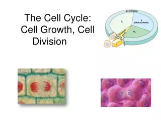
Phases of Cell cycle • Cell cycle involves two major phases S phase in which DNA duplication takes place (S for synthesis of DNA), takes about half time (10-12 hours) of cell cycle and M phase in which mitosis takes place (takes about one hour). • Most cells require much more time to grow and double their mass of proteins and organelles than they require to replicate their DNA and divide. To allow more time for growth, extra gap phases are inserted in most cell cycles a G1 phase between M phase and S phase and a G2 phase between S phase and mitosis. • So the eukaryotic cell cycle is divided into four sequential phases: G1, S, G2, and M. • G1, S, and G2 together are called interphase. In a typical human cell, interphase might occupy 23 hours of a 24 hour cycle, with 1 hour for M phase. • If extra cellular conditions are unfavorable, for example, cells delay progress through G1 and may even enter a specialized resting state known as G0 (G zero), in which they can remain for days, weeks, or even years before resuming proliferation.
Figure 17-3. The phases of the cell cycle. The cell grows continuously in interphase, which consists of three phases: DNA replication is confined to S phase; G1 is the gap between M phase and S phase, while G2 is the gap between S phase and M phase. In M phase, the nucleus and then the cytoplasm divide.
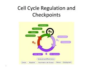
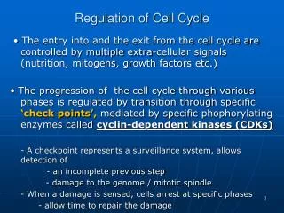
The Control System Can Arrest the Cell Cycle at Specific Checkpoints • In most cells there are several points in the cell cycle, called checkpoints, at which the cycle can be arrested if previous events have not been completed, for example if DNA replication is not properly takes place , the cell will not enter in the mitosis stage.
Figure 17-33. How DNA damage arrests the cell cycle in G1. When DNA is damaged, protein kinases that phosphorylate p53 are activated. Mdm2 normally binds to p53 and promotes its ubiquitylation and destruction in proteasomes. Phosphorylation of p53 blocks its binding to Mdm2; as a result, p53 accumulates to high levels and stimulates transcription of the gene that encodes the CKI protein p21. The p21 binds and inactivates G1/S-Cdk and S-Cdk complexes, arresting the cell in G1. In some cases, DNA damage also induces either the phosphorylation of Mdm2 or a decrease in Mdm2 production, which causes an increase in p53 (not shown).
The Cell-Cycle Control System Is Based on Cyclically Activated Protein Kinases • Cell-cycle control system is a family of protein kinases known as cyclin-dependent kinases (Cdks). • The activity of these kinases rises and falls as the cell progresses through the cycle. • An increase in Cdk activity at the beginning of mitosis, for example, leads to increased phosphorylation of proteins that control chromosome condensation, nuclear envelope breakdown, and spindle assembly. • Cyclical changes in Cdk activity are controlled by a complex array of enzymes and other proteins. The most important of these Cdk regulators are proteins known as cyclins. When cyclin binds with cdk they become active and have kinase activity. • Cyclins were originally named as such because they undergo a cycle of synthesis and degradation in each cell cycle.
Figure 17-16. A simplified view of the core of the cell-cycle control system. Cdk associates successively with different cyclins to trigger the different events of the cycle. Cdk activity is usually terminated by cyclin degradation. For simplicity, only the cyclins that act in S phase (S-cyclin) and M phase (Mcyclin) are shown, and they interact with a single Cdk; as indicated, the resulting cyclin-Cdk complexes are referred to as S-Cdk and M-Cdk, respectively.
Figure 17-17. The structural basis of Cdk activation. (A) In the inactive state, without cyclin bound, the active site is blocked by a region of the protein called the T-loop (redof cyclin causes). (B) The binding the T-loop to move out of the active site, resulting in partial activation of the Cdk2. (C) Phosphorylation of Cdk2 (by CAK) at a threonine residue in the T-loop further activates the enzyme by changing the shape of the T-loop, improving the ability of the enzyme to bind its protein substrates.
Checkpoints Generally Operate Through NegativeIntracellular Signals • Checkpoint mechanisms operate through negative intracellular signals that arrest the cell cycle, rather than through the removal of positive signals that normally stimulate cell-cycle progression.
Cdk Activity Can Be Suppressed Both by Inhibitory Phosphorylation and by Inhibitory Proteins Figure 17-18. The regulation of Cdk activity by inhibitory phosphorylation. The active cyclin-Cdk complex is turned off when the kinase Wee1 phosphorylates two closely spaced sites above the active site. Removal of these phosphates by the phosphatase Cdc25 results in activation of the cyclin-Cdk complex. For simplicity, only one inhibitory phosphate is shown. The activating phosphate is added by CAK, as shown in Figure 17-17.
Figure 17-19. The inhibition of a cyclin-Cdk complex by a CKI. This drawing is based on the three-dimensional picture of the human cyclin ACdk2 complex bound to the CKI p27, as determined by x-ray crystallography. The p27 binds to both the cyclin and Cdk in the complex, distorting the active site of the Cdk. It also inserts into the ATP-binding site, further inhibiting the enzyme activity.
The Cell-Cycle Control System Depends on Cyclical Proteolysis Figure 17-20. The control of proteolysis by SCF and APC during the cell cycle. (A) The phosphorylation of a target protein, such as the CKI shown, allows the protein to be recognized by SCF, which is constitutively active. With the help of two additional proteins called E1 and E2, SCF serves as a ubiquitin ligase that transfers multiple ubiquitin molecules onto the CKI protein. The ubiquitylated CKI protein is then immediately recognized and degraded in a proteasome.
(B) M-cyclin ubiquitylation is performed by APC, which is activated in late mitosis by the addition of an activating subunit to the complex. Both SCF and APC contain binding sites that recognize specific amino acid sequences of the target protein.
Figure 17-23. The activation of M-Cdk. Cdk1 associates with M-cyclin as the levels of M-cyclin gradually rise. The resulting M-Cdk complex is phosphorylated on an activating site by the Cdk-activating kinase (CAK) and on a pair of inhibitory sites by the Wee1 kinase. The resulting inactive M-Cdk complex is then activated at the end of G2 by the phosphatase Cdc25. Cdc25 is stimulated in part by Polo kinase, which is not shown for simplicity. Cdc25 is further stimulated by active M-Cdk, resulting in positive feedback. This feedback is enhanced by the ability of M-Cdk to inhibit WeeI.
Figure 17-26. The triggering of sister-chromatid separation by the APC. The activation of APC by Cdc20 leads to the ubiquitylation and destruction of securin, which normally holds separase in an inactive state. The destruction of securin allows separase to cleave a subunit of the cohesin complex holding the sister chromatids together. The pulling forces of the mitotic spindle then pull the sister chromatids apart. In budding yeasts at least, cohesin cleavage by separase is facilitated by the phosphorylation of the cohesin complex adjacent to the cleavage site, just before anaphase begins. The phosphorylation is mediated by Polo kinase and provides an additional control on the timing of the metaphase-to-anaphase transition.
Summary Figure 17-34. An overview of the cell-cycle control system. The core of the cellcycle control system consists of a series of cyclin-Cdk complexes (yellow). The activity of each complex is also influenced by various inhibitory checkpoint mechanisms, which provide information about the extracellular environment, cell damage, and incomplete cell-cycle events (top).