Download
1 / 67
700 likes | 827 Vues
This chapter delves into seminal experiments and findings that shaped our understanding of DNA as the genetic material. It covers Frederick Griffith's bacterial transformation in 1928 using Streptococcus pneumoniae, Avery, MacLeod, and McCarty's identification of DNA as the transforming agent, and Hershey & Chase's groundbreaking Blender Experiment in 1952. The narrative culminates in Watson and Crick’s revelation of DNA's double helix structure, built upon previous work by Chargaff, Wilkins, and Franklin. This journey highlights the collaborative nature and complexity of scientific discovery in genetics.

E N D
DNA - The Code of Life Chapter 16
F. Griffith & Transformation • 1928 • Streptococcus pneumoniae • 2 strains: R – harmless, S – pathogenic • Mixed inactive S strain & active R strain; injected into mouse • Mouse died, pathogenic strain in blood • Transformation
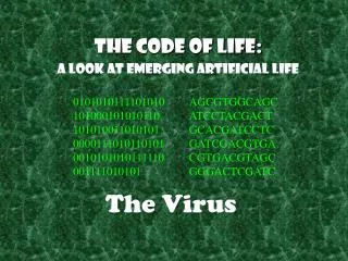
Avery, McCarty & MacLeod • 1944 : transforming agent is DNA • Skeptics – bacteria not complex • More research • Viruses (bacteriophages) • Viral structure & replication?
Hershey & Chase – The Blender Experiment • 1952: DNA is the genetic material of the phage T2 • T2 phage infects E. coli • Labeled protein coat with radioactive S and the DNA with radioactive P • Phages infect E. coli separately • Only P found in the bacterium
Findings • Infected with radio-labeled proteins - radioactivity in supernatant • Infected with radio-labeled DNA - radioactivity in pellet • Hershey & Chase’s conclusion?
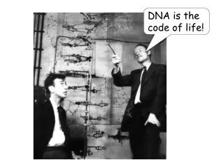
Let the race begin! • 1950s – scientific community racing to find 3-D structure of DNA • Major players • James Watson & Francis Crick • Linus Pauling • Maurice Wilkins & Rosalind Franklin
Puzzle Pieces • Chargaff’s Data • Backbone Structure – single strand • Franklin’s Data
Chargaff’s Rules - Structure • 1947 • Polymer - deoxyribose sugar, phosphate grp & nitrogen-containing base • Bases: • adenine (A), thymine (T), guanine (G), or cytosine (C)
Chargaff’s Rules • Certain bases were always equal in number • # adenines approx equal to # of thymines (%T = %A) • # guanines approx equal to # of cytosines (%G = %C) • Why is this significant?
Backbone Structure • Phosphate group of one nucleotide attached to the sugar of the next • Result is a “backbone” of phosphates and sugars, from which the bases project
R. Franklin & M. Wilkins X-Ray Crystallography
Franklin’s Data • Wilkins & Franklin used X-ray crystallography to study DNA structure • X-rays diffracted as they pass through aligned fibers of purified DNA • Diffraction pattern used to deduce 3-D shape of molecules
What the picture tells us • DNA was helical in shape!
Putting the puzzle together… • Watson & Crick – “stolen” ideas or a product of the times? • However… it was Watson who deduced the width of the helixand the spacing of bases • Model building to beat Pauling
TRIAL AND ERROR • Watson & Crick began to work on a model of DNA with two strands, the double helix • Wire molecular models - first tried to place the sugar-phosphate chains on the inside • Did not fit the X-ray measurements and other info on chemistry of DNA
Breakthrough • Watson put sugar-phosphate chain on the outside & nitrogen bases on the inside
Nuts & Bolts – Chargaff’s Data • Watson & Crick determined that chemical side groups off nitrogen bases formed H bonds, connecting strands • Adenine - form 2 H bonds only with thymine • Guanine - form 3 H bonds only with cytosine • Findings explained Chargaff’s rules
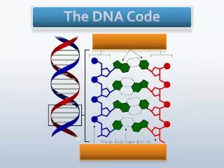
DNA Structure • DNA bases: • Purines: adenine and guanine • Pyrimidines: thymine and cytosine • Nucleotides are covalently bonded with a sugar-phosphate backbone • The linkage forms a 3’,5’ phosphodiester linkage • One end of the molecule has a free 5’ carbon; the other has a free 3’ carbon
DNA Structure • Two polynucleotide chains intertwined to form a double helix
Technical DNA Data • .34 nm is the distance between the bases • 3.4 nm repeat of nucleotides due to a complete “turn” of the helix • width of molecule is 2.0 nm
Pyrimidines and Purines • Pyrimidines are single-ringed • Purines are double-ringed • Bonding is complementary • Sequence in one chain dictates sequence in opposite chain
DNA Replication • Complimentary bases act as templates • Bases of one strand allow for exact duplication
DNA Replication • Origin(s) of replication • Prokaryotes - single specific sequence of nucleotides recognized by replic. enzymes • Replication proceeds in both directions • Eukaryotes – hundreds/thousands of origin sites per chrom • Bubble with replication forks at each end • Bubbles elongate as DNA is replicated and eventually fuse
Bidirectional Synthesis • In prokaryotes, the circular DNA is opened up, and synthesis occurs in both directions
Bidirectional Synthesis • In eukaryotes, the linear DNA has many replication forks
DNA Replication • Proteins and enzymes work together • DNA strands must be unwound during replication • DNA helicase unwinds the strands • Single stranded binding proteins (SSB) prevent immediate reformation of the double helix • Topoisomerases break and then rejoin the strands, “untying” the knots that form
DNA Replication Order • Always proceeds in a 5’ 3’ direction • DNA polymerase can add only at the 3’ end • Nucleotides are polymerized and 2 phosphates are removed in the process
Nucleotide Synthesis • Raw nucleotides are nucleoside triphosphates • N base, deoxyribose, & a triphosphate tail • Nucleotide added, last 2 phosphate grps hydrolyzed, forming pyrophosphate • Exergonic rxn drives polymerization of the nucleotide to the new strand
DNA Pol • DNA polymerases can only add nucleotides to the free 3’ end of a growing DNA strand • A new DNA strand can only elongate in the 5’ 3’ direction
Problems? • One parental strand is oriented 3’ 5’ into the fork, while the other is oriented 5’ 3’ into the fork • At fork, only one parental strand (3’ 5’ into the fork), leading strand, can be used by polymerases as a template for a continuous complementary strand
Continuous & Discontinuous • Replication is continuous on one strand and discontinuous on the other • Replication begins at replication forks
Okazaki fragments • Synthesis of the leading strand is continuous • The lagging strand (discontinuous) is synthesized in pieces called Okazaki fragments
Okazaki fragments • 100 - 1000 nucleotides in length • Initiated by a separate RNA primer • Okazaki fragments are joined together by DNA ligase
RNA Primer • DNA pol cannot initiatesynthesis because it can only add nucleotides to end of an existing chain • Requires an RNA primer • Primase, an RNA pol, links ribonucleotides complementary to the DNA template into the primer • RNA pol can start an RNA chain from a single template strand