Download
1 / 101
1.27k likes | 3.59k Vues
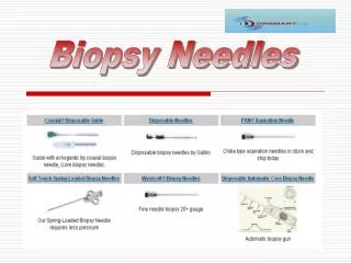
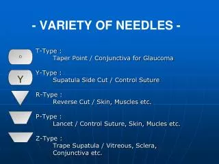
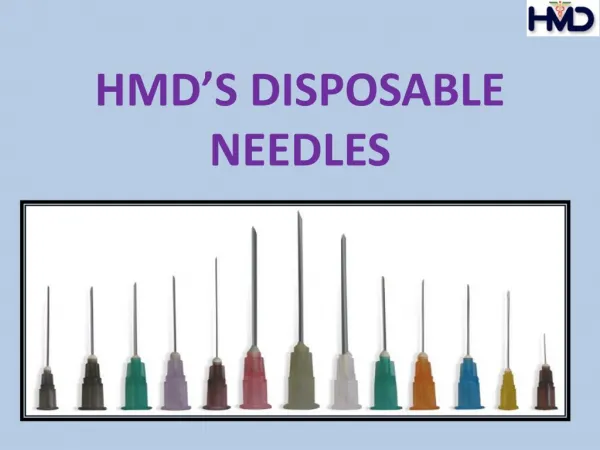
Types of needles. Curved needles. 1/2 of circle. 3/8 of circle. 5/8 of circle. Types of needles according to the threading. Eyed. Eyeless. Types of needles according to cut section. Cutting needle. Rounded needle. Control of 1ry hemorrhage. 1ry hemorrhage. Place pack for 4 min.

E N D
Curved needles 1/2 of circle 3/8 of circle 5/8 of circle
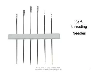
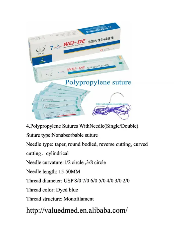
Types of needles according to the threading Eyed Eyeless
Types of needles according to cut section Cutting needle Rounded needle
1ry hemorrhage Place pack for 4 min
3 ligations 2 ligations Artery :high pressure Vein :low pressure
Tension suture It takes muscles &peritoneum On tension of the suture
Sit of the incision Mid axillary line Ant. Axillary line
Med. axillary line Ant. axillary line Points of drain
Lung Spread the muscle fibers by artery forceps to expose pleura
Pierce pleura and introduce finger to confirm entry to pleura
Make sure that the incision is large enough to accommodate the drain + your finger Drain
Rib Drain Rib
Stomach Gastrosplenic lig. Ant.lienorenal lig Spleen Pancreas Kidney Post.lienorenal lig.
Gastro pherenic ligament Spleen Anterior lienorenal ligament Pancreas posterior lienorenal ligament
Gastropherenic ligament Ant. layer of lienorenal ligament Post. Layer of lienorenal ligament
Stomach (medially) Spleen (medially) Hot packs Divide the posterior layer of lienorenal ligament
Divide the posterior layer of lienorenal ligament Traction of the spleen medially
Ant. Lienorenal lig. Pancreas Splenic artery Spleen Pos. lienorenal lig.
Squeeze the spleen Divide splenic vein
Rectum Anal canal Overall view of the rectum and anal canal
Anatomy of rectum and anal canal Rectum Anal canal Dentate line External sph. Internal sph.
Superior rectal vein Internal plexus of veins External anal sphincter Anal canal Internal anal sphincter Anatomical anal canal structure
Internal opening of the fistula External opening of the fistula The track of the fistula
Insertion of probe Probe