HKIN 473
HKIN 473. Recording Motor Units. Recording Electrical Signals. Muscle fiber sarcolemma action potential is very small ~ 1 millivolt. Therefore, to be able to record and “see” muscle activity using indwelling electrodes, need to amplify the signal first. 1) Preamplifier 2) Amplifier

HKIN 473
E N D
Presentation Transcript
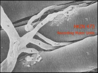
HKIN 473 Recording Motor Units
Recording Electrical Signals • Muscle fiber sarcolemma action potential is very small ~ 1 millivolt. • Therefore, to be able to record and “see” muscle activity using indwelling electrodes, need to amplify the signal first. • 1) Preamplifier 2) Amplifier • For our laboratory, need to amp Motor unit signal by between 1,000 and 5,000 times. • Gain = Output/Input (1 Volt = 1,000/1 mv)
Sampling Signals to Computer • Sample signals from Muscle Amp to computer using A/D (analog to digital) converter.
Sampling Signals to Computer • Need to sample at the right frequency to see the detail of the signal. • ~10,000 Hzfor MU recordings in humans • To do this, need to know: 1) the frequency (how many times a second) an event of interest occurs (1, 10, 100 times a second?). 2) the detail of the signal you are sampling. If you do not sample (look at signal) frequently enough may miss events or not get a clear enough picture of the real signal. In MU recordings we use shape of the potential or its morphology to identify it as a single recording.
Recording Motor Units • Important Issues When Analyzing Motor Units: • Single Unit Recordings • Pay Attention to One Unit • Sampling Issues • Motor Unit Morphology (Shape) • This is done using both visual signals (oscilloscope) as well as audio signals (units may have different sounds)
External and Internal Stimuli Sensory Systems Vision Higher Centers Vestibular Motor Systems Somatosensory Spinal Cord Motoneurons Sensory Feedback Muscles Motor Output
External and Internal Stimuli Sensory Systems Vision Higher Centers Vestibular Motor Systems Somatosensory Spinal Cord Kennedy and Cresswell 2001 Inglis et al. 2002 Kennedy and Inglis 2001 Motoneurons Sensory Feedback Muscles Motor Output
External and Internal Stimuli Sensory Systems Vision Higher Centers Vestibular Motor Systems Somatosensory Spinal Cord Kennedy et al. (Submitted) Kennedy et al. (In Press) Kennedy and Inglis 2002 Kennedy and Inglis 2001 Motoneurons Sensory Feedback Muscles Motor Output
2 mV Test Reflex 10 ms Galvanic Stimulation and Muscle Reflexes Cathode-Forward Cathode-Left Anode-Forward Anode-Right Conditioned Reflex
Threshold Force 200 N Neurogram 1.4 mV 4 s Anode Cathode No Stimulus
Environment Motor Response Sensory Signals CNS Senses Effectors
Microneurography Set-up • Subject comfort • Reference Electrode • Recording Electrode • Muscle Activity (EMG) • Force Probe
Microneurography Explored • Needle manually placed in a peripheral nerve • Needle can record from nearby axons • Shape of action potentials dependent upon position of needle
Maintained Indentation Fast Adapting Type I Fast Adapting Type II Slow Adapting Type I Slow Adapting Type II
Skin Receptors in the Foot Sole Location of the Receptive Fields Receptors 104 SA Type I 15 (14%) SA Type II 16 (15%) FA Type I 59 (57%) FA Type II 14 (14%)
Skin Receptors in the Foot Sole • Slow adapting code • Base of support • Joint position Base of Support Surface Contact Slip Center of Pressure Slip • Fast adapting code • Movement of the center of pressure • Detection of slip