White Blood Cell Imaging in Diagnostic Applications
130 likes | 230 Vues
Explore the diagnostic applications of labeled white blood cells using 111In and 99mTc in various medical conditions such as abscess evaluation, osteomyelitis detection, and more.

White Blood Cell Imaging in Diagnostic Applications
E N D
Presentation Transcript
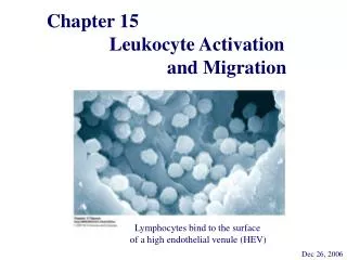
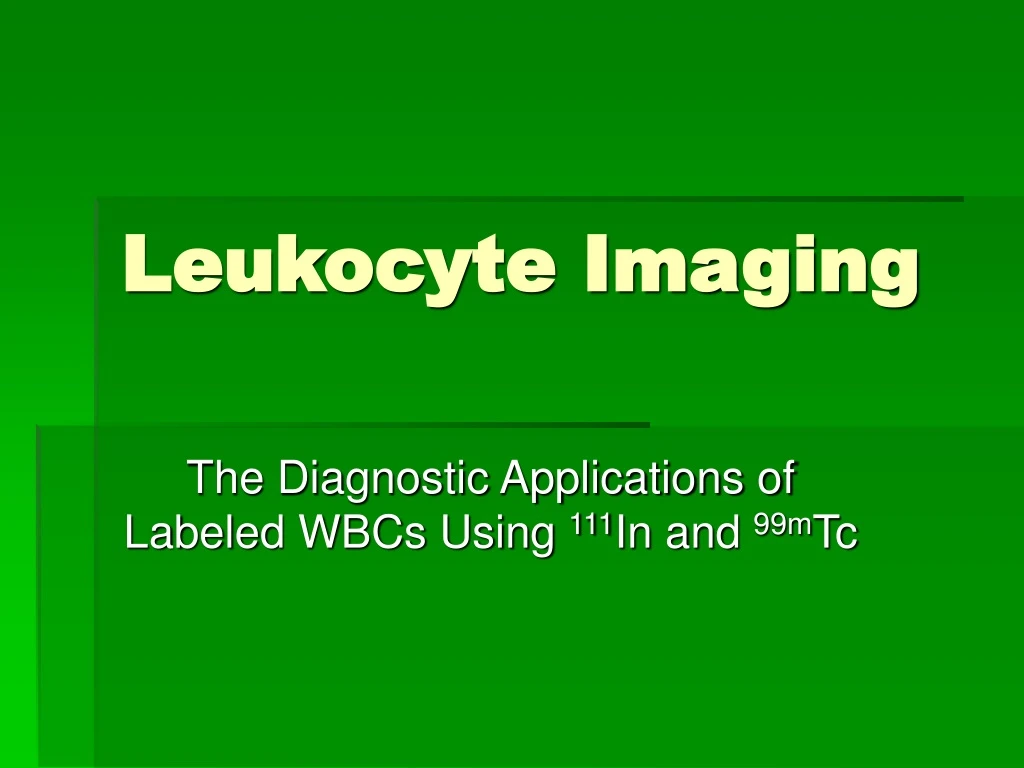
Leukocyte Imaging The Diagnostic Applications of Labeled WBCs Using 111In and 99mTc
Normal 111In WBC Scan • The patient is status post left hemicolectomy with intra-abdominal fluid collection. Evaluate for abscess. • FindingsWhite blood cell activity is seen in the spleen, liver, and axial skeleton, as expected. No areas of abnormally increased white cell activity are seen. • ConclusionsNormal examination with no evidence for abscess.
99mTcMDP + 111IN-WBC • Left Above: 99mTc MDP Bone Scan indicates increased activity in the proximal tibia and patella. Osteomyelitis (bone infection) cannot be ruled out. • Left Below: 111Indium-Oxine White Blood Cell Label of same patient reveals an area of increased activity indicating an infection in the left proximal tibia and distal femur. • Images courtesy of Nuclear Medicine Department, Dartmouth-Hitchcock Medical Center
111Indium WBC Imaging WBC study shows increased WBC activity in the L femur at site of plate attachment. This uptake correlates with evidence of hyperemia seen on both the sulfur colloid study and the blood pool phase of the bone scan. These findings are strongly suggestive of cellultis in the soft tissues lateral to the left femoral plate. No other abnormal foci of white blood cell accumulation are identified within the chest, abdomen, or pelvis. • Clinical History84-year-old man with left lower extremity and hip pain.
111Indium WBC Scan • Clinical HistorySevere mitral regurgitation and cardiac valvular surgery. FUO; ? abscess. The study demonstrates three foci of abnormal white blood cell activity. One is in the region of the heart, another in the medial right lower chest cavity and the third in the right upper chest cavity. Correlation with chest radiograph is recommended for further delineation of chest findings. Infected valve and endocarditis should be excluded.
99mTc HMPAO Labeled WBC • Superior to In111 Leukocyte labeling, due to readily available and cheaper. • Best for osteomyeolitis in the extremities • Higher counting statistics and can block pelvic area due to excretion thru urine. • Imaging time is decreased from 10 minutes to 5 minutes. • Can Imaging within hours not 24 hours • SPECT Imaging can be performed
Tc99m HMPAO WBC Scan • Tc-99m-labeled WBC Scan obtained one hour after injection revealed abnormal patchy uptake throughout the bone marrow. Normal hepatic and splenic uptake. No collections of tracer to suggest a focus of infection outside the reticuloendothelial system. Residual bowel activity in the right lower quadrant of the abdomen due to the Tc-99m-DISIDA hepatobiliary scan performed 20 hours earlier was noted prior to reinjection of the autologous labeled white cells.
Tc99m Leukocyte Scan The image on the left shows a technetium labeled white blood cell scan of the feet of a diabetic patient with infection in the great toe of the left foot. Notice the dark spot at the site of infection.
99mTcWBC Scan • Male Patient 1 month post distal pancreatectomy and splenectomy. The patient was febrile at time of study. • Images consistent with inflammation in surgical wound and pelvic abscess. LUQ Incision Pelvic Abscess
Inflammatory Bowel With 99mTcWBC 24 Year old female, known IBD • Diffuse uptake throughout the colon. No small bowel uptake identified. • When the disease involves the entire colon and there is no small bowel activity, total colectomy may be considered.
99mTc WBC Liver Abscess 48 Year old male, FUO and abdominal pain. • WBC Scan demonstrates 5 abscesses. • 4 located in the right lobe of the liver and a single abscess was noted in the left (quadrate) lobe of the liver.
In111 Leukocytes Dose 200-500uCi Image 10 minutes per view 20% window Medium or High energy collimator Dual energies 173 keV and 247 keV Normal uptake spleen, Liver and bone marrow 0-4 hours post injection shows transient pulmonary distribution Image 24 hours post injection Possibly 3-4 hours for abdominal imaging Tc99m Ceretec Leukocytes Dose 20-25 mCi Image 5 minutes per view 20% window Low energy-high resolution Energy setting 140keV Normal uptake: liver, spleen, bone marrow, kidneys and bladder Post 4 hours intestinal excretion may be evident Image at 1-4 hours post injection In111 Vs. Tc99m