Download
1 / 22
240 likes | 1.38k Vues
Diagnosis of Acute Ischemic and Hemorrhagic Stroke. Ischemic Stroke. Low blood flow to focal part of brain Usually caused by thromboembolism Acute therapy includes thrombolysis 2 prevention depends on source of thromboembolus Accounts for 85% of strokes.

E N D
Ischemic Stroke • Low blood flow to focal part of brain • Usually caused by thromboembolism • Acute therapy includes thrombolysis • 2 prevention depends on source of thromboembolus • Accounts for 85% of strokes
Transient Ischemic Attack (TIA) • Reversible focal dysfunction, usually lasts minutes • Among TIA pts who go to ED: • 5% have stroke in next 2 days • 25% have recurrent event in next 3 months • Stroke risk decreased with proper therapy
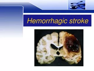
Intracerebral Hemorrhage • Bleeding into brain tissue • Usually caused by chronic hypertension • Non-hypertension cause more likely if: • No past history of hypertension • Lobar (i.e., peripheral, not subcortical) • May require emergency surgery • Accounts for 10% of strokes
Subarachnoid Hemorrhage • Bleeding around brain • Usually caused by ruptured aneurysm • Surgical emergency • Cerebral angiography • Aneurysmal clipping • Accounts for 5% of strokes
Five Major Stroke Syndromesfor Rapid Recognition in the ED All Occur Suddenly in Stroke Patients • Left (dominant) cerebral hemisphere • Right (nondominant) cerebral hemisphere • Brainstem • Cerebellum • Hemorrhage Note: The dominant cerebral hemisphere is the side that controls language function.
Left (Dominant)Cerebral Hemisphere • Aphasia • L gaze preference • R visual field deficit • R hemiparesis • R hemisensory loss
Right (Nondominant)Cerebral Hemisphere • Neglect (= L hemi-inattention) • R gaze preference • L visual field deficit • L hemiparesis • L hemisensory loss
Brainstem • Hemi- or quadriparesis • Sensory loss in hemibody or all 4 limbs • Crossed signs (face 1 side, body other side) • Diplopia, dysconjugate gaze, gaze palsy • Vertigo, tinnitus • Nausea, vomiting • Hiccups, abnormal respirations • Decreased consciousness
Cerebellum • Truncal = gait ataxia • Limb ataxia
Hemorrhage Symptoms only suggestive of hemorrhage. CT or LP needed for definitive diagnosis. • Headache • Neck stiffness • Neck pain • Light intolerance • Nausea, vomiting • Decreased consciousness
Acute Stroke ScalesMost Commonly Used in the U.S. • Glasgow Coma Scale ( LOC) • Hunt & Hess Scale (SAH) • NIH Stroke Scale (AIS)
Glasgow Coma ScaleAdd the 3 scores (1 from each category) Best Verbal 5 oriented 4 confused 3 inappropriate 2 incomprehensible 1 none Eye Opening 4 spontaneous 3 to speech 2 to pain 1 none Best Motor 6 obeys commands 5 localizes pain 4 withdraws to pain 3 abnl flexion to pain 2 extension to pain 1 none Quantifies deficits in pt w/ LOC: GCS < 9 carries poor prognosis
Hunt and Hess ScaleChoose the single-most-appropriate grade • Grade I: asx; mild HA; slight nuchal rigidity • Grade II: moderate-to-severe HA; nuchal rigidity; • no neuro deficit other than CN palsy • Grade III: drowsiness/confusion; mild focal deficit • Grade IV: stupor; moderate-to-severe hemiparesis • Grade V: coma; decerebrate posturing • Prognostic value in SAH pts: • Grades I-III better prognosis & surgical candidates
Urgent Evaluation of Patients with Focal Neurologic Deficits • Complete neurologic exam • lengthy, variable, parts not reproducible • inappropriate in acute setting • Glasgow Coma Scale • valuable for pts w/ LOC • does not quantify focal neurologic deficit • Hunt & Hess Scale • value is specific to SAH pts
NIH Stroke Scale • Designed for acute ischemic stroke trials • Relatively quick (5-10 min) and reproducible • Requires speech-&-language cards, safety pin, complex grading scale • Quantifies stroke deficit: < 4 = mild stroke > 15 = poor prognosis if no treatment > 22 = risk for intracranial hemorrhage after t-PA
Mental Status LOC Questions Commands Language Neglect Cranial Nerves Visual fields Horizontal gaze Face strength Dysarthria NIH Stroke Scale:Modified arrangement of items • Limbs • R/L arm motor • R/L leg motor • Coordination • Sensation
1a. LOC 1b. LOC questions 1c. LOC commands 2. Best gaze 3. Visual fields 4. Facial palsy 5a. Right arm motor 5b. Left arm motor 6a. Right leg motor 6b. Left leg motor 7. Limb ataxia 8. Sensory 9. Best language 10. Dysarthria 11. Extinction/ inattention NIH Stroke Scale:“Traditional” order of items
NIH Stroke Scale:Caveats re: “traditional” version • Item 12—Distal Motor Function • was never included in total NIHSS score • is supplemental and not necessary • Grades of “9”—Untestable • used only for motor, ataxia, and dysarthria • number 9 assigned for computer purposes • do NOT give 9 points for untestable items
Stroke Differential Diagnosis:Sudden Onset Persistent Focal Deficit • Ischemic stroke • Intracerebral hemorrhage • Partial seizure with postictal (Todd’s) paralysis • Abscess with seizure • Tumor with bleed or seizure • Toxic-metabolic insult with old cerebral lesion • Hypoglycemia • Subdural hematoma (acute) • Multiple sclerosis • Cerebritis
Stroke Differential Diagnosis:Sudden Onset Transient Focal Deficit • Transient ischemic attack • Partial seizure • Migraine with aura NOTE: AVMs can cause all three types of transient focal neurologic deficits.
Persistent LOC Subarachnoid hemorrhage Meningitis Drug overdose Toxic-metabolic insult Seizure with postictal state Subclinical status epilepticus Transient LOC Seizure Syncope Stroke Differential Diagnosis:Depressed LOC without Focal Deficit