Download
1 / 14
200 likes | 300 Vues
Learn about the anatomy of the vitreous humor, conditions like synchisis and vitreous hemorrhage, causes, symptoms, and treatment options. Explore how vitreous opacities impact vision and the clinical significance of vitreous disorders.

E N D
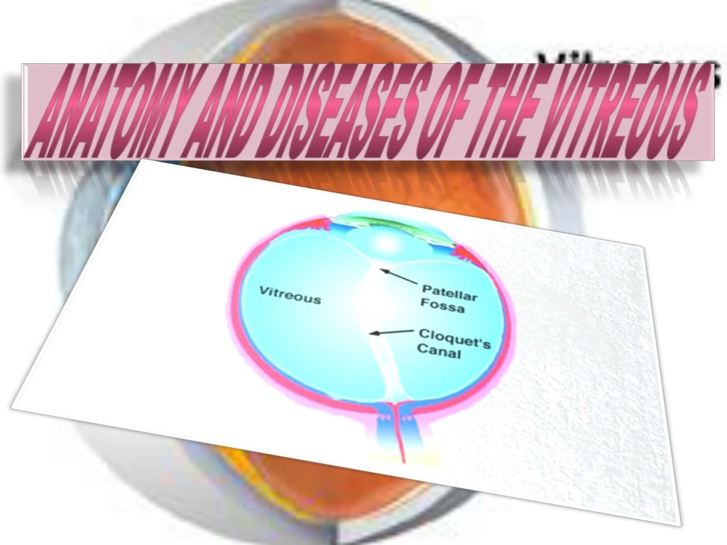
Anatomy of the vitreous The vitreous humor is a transparent jelly like structure which helps to maintain the shape of the eyeball and optical function .it has all the properties of the hydrophilic gel, undergoing turgescence. It contains a few cells and an obliterated canal known as hyaloids canal it has no blood vessels. Its surface is condensed to form thin membrane called the hyaloids membrane.
Diseases of the vitreous Fluidity of vitreous (synchisis)- it is due to conversion of the colloidal gel into a sol Causes: 1)Senility 2)Degenerative myopia 3)Uveitis 4)Trauma-concussion or intraocular foreign body 5)Treatment of retinal detachment by diathermy or light coagulation
Clinical significance: 1)Development of retinal detachment 2)During lens extraction-posterior dislocation of lens and vitreous loss may occur. That is why in cases of degenerative myopia, planned extracapsular lens extraction is indicated.
Vitreous opacities: Subjectively they are seen entoptically as muscaevolitantes. Any opacity in the eye which casts a shadow upon the sentient elements of the retina (rods and cones) will appear as dark spot in the field of vision. However, an opacity in the retinal blood vessels (RBCs) will not produce muscae
Causes: • Congenital remnants of the hyaloid vascular system. • Endogenous opacities • Coagula of the colloid basis of the gel- senility, degenerative myopia. • Crystalline deposits- asteroid bodies, synchisis scintillans. • Exogenous opacities
Exogenous opacities • Protein coagula- plasmoid vitreous as in iridocyclitis and choroiditis. • Exudative cells. • Blood. • Tissue cells-epithelial cells, histocytes. • Tumors cells. • Pigment-melanin, haemosiderin .
Symptoms: • Muscae volitantes may appear in the form of black dots, lines or cobwebs. • When many vitreous opacities are present there may be haziness of vision. Vision is often best in the morning before the vitreous has been stirred up by movements of the eye. • In a myopia sudden increase in muscae volitantes may herald the onset of retinal detachment.
Signs: • The opacities may be seen: • With the slit lamp. • As black opacities moving independently of the movements of the eye by distant direct ophthalmoscopy. • With an ophthalmoscope with a +10 D lens.
Treatment: • The patient should be advised to ignore the opacities as much as possible. • Treatment of the underlying cause –iridocyclitis-choroiditis. • Vitrectomy in desperate cases.
Vitreous hemorrhage: Source: Retinal blood vessel-veins or arteries. Vessels of the ciliary body. New vessels growing into vitreous-retinitis proliferans . Spilling over of hyphaema after cataract extraction.
Causes: • Trauma. • Diabetes mellitus with proliferative retinopathy. • Hypertension. • Eales’ disease. • Blood dyscrasias - anemia, hemophilia, sickle cell disease.
Small vitreous hemorrhage may get absorbed but in recurrent hemorrhages retinitis proliferans may develop. • Treatment: • Photocoagulation in cases of diabetic retinopathy. • Vitrectomy.