Download
1 / 77
770 likes | 945 Vues
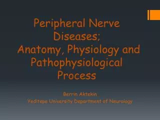
Peripheral Nerve Diseases; Anatomy, Physiology and Pathophysiological Process. Berrin Aktekin Yeditepe University Department of Neurology. Functional Organization of the PNS. Figure 14.1. Basic Anatomical Scheme of the PNS in the Region of a Spinal Nerve.

E N D
Peripheral Nerve Diseases; Anatomy, Physiology and Pathophysiological Process Berrin Aktekin Yeditepe University Department of Neurology
Functional Organization of the PNS Figure 14.1
Basic Anatomical Scheme of the PNS in the Region of a Spinal Nerve A nerve is composed of numerous nerve fibers Figure 14.2
Spinal Nerves • 31 pairs – contain thousands of nerve fibers • Connect to the spinal cord • Named for point of issue from the spinal cord - 8 pairs of cervical nerves (C1 – C8) - 12 pairs of thoracic nerves (T1 – T12) - 5 pairs of lumbar nerves (L1 – L5) - 1 pair of coccygeal nerves (Co1)
Spinal Nerves • Connect to the spinal cord by the dorsal root and ventral root • Dorsal root – contains sensory fibers - cell bodies located in the dorsal root ganglion • Ventral root– contains motor fibers arising from anterior gray column
Spinal Nerves • Branch into dorsal ramus and ventral ramus - dorsal and ventral rami contain sensory and motor fibers • Rami communicantes – connect to the base of the ventral ramus - lead to the sympathetic chain ganglia
Innervation of the Back • Dorsal rami – innervate back muscles - follow a neat, segmented pattern - innervate a horizontal strip of muscle and skin (in line with emergence point from the vertebral column)
Innervation of the Back Figure 14.7b
Innervation of the Anterior Thoracic and Abdominal Wall • Thoracic region – ventral rami arranged in simple, segmented pattern • Intercostal nerves supply intercostal muscles, skin, and abdominal wall - each gives off lateral and anterior cutaneous branches
Introduction to Nerve Plexuses • A network of nerves • Ventral rami (except T2 – T12) - branch and join with one another - form nerve plexuses in the cervical, brachial, lumbar, and sacral regions - primarily serve the limbs - fibers from ventral rami crisscross
The Cervical Plexus • Buried deep in the neck under the sternocleidomastoid muscle • Formed by ventral rami of first 4 cervical nerves (C1 – C4) • Most are cutaneous nerves • Some innervate muscles of the anterior neck • Phrenic nerve – major nerve
The Brachial Plexus and Innervation of the Upper Limb • Brachial plexus lies in the neck and axilla • Formed by ventral rami of C5 – C8 • Cords give rise to main nerves of the upper limb
Fig 14.9a The Brachial Plexus
Lumbar Plexus and Innervation of the Lower Limb • Arises from L1 – L4 • Smaller branches innervate the posterior abdominal wall and psoas muscle • Main branches innervate the anterior thigh - femoral nerve innervates anterior thigh muscles - obturator nerve innervates adductor muscles
Sacral Plexus • Arises from spinal nerves L4 – S4 • Caudal to the lumbar plexus • Often considered with the lumbar – lumbosacral plexus • Sciatic nerve – largest nerve - 2 nerves in one sheath: Tibial nerve – innervates most of the posterior lower limb; Common fibular (peroneal) nerve – innervates muscles of the anterolateral leb
Autonomic Nervous System • General visceral motor part of the PNS • Has 2 divisions (with opposite effects): - Parasympathetic: ‘housekeeping’ activities (rest and digest) - Sympathetic: extreme situations (fight or flight)
Sensory System • Five sense !!! • Peripheral Sensory System • Spinothalamic • Dorsal Column • Cortical-integrative Sensory System • Visceral Sensory System
Innervation of the Skin: Dermatomes • Dermatome – an area of skin • Innervated by cutaneous branches of a single spinal nerve • Upper limb – skin is supplied by nerves of the brachial plexus • Lower limb: Lumbar nerves – anterior surface Sacral nerves – posterior surface
Peripheral Sensory System • Spinothalamicsystem-Cutaneous • Pain- • Temperature • Light touch/pressure • Dorsal Column-Medial Lemniscal System-Proprioception • Vibration • Position
Peripheral Sensory Receptors • Most fit into 2 main categories: 1. free nerve endings of sensory neurons - monitor general sensory information such as touch, pain, pressure, temperature, and proprioception 2. complete receptor cells – specialized epithelial cells or small neurons that transfer sensory information to sensory neurons - monitor most special sensory information such as taste, vision, hearing, and equilibrium
Sensory Receptors of the PNS • Also classified according to: a) Location – based on body location or location of stimuli to which they respond b) Type of stimulus detected – kinds of stimuli that most readily activate them c) Structure – divided into 2 broad categories free or encapsulated nerve endings
Classification by Location • Exteroceptors – sensitive to stimuli arising from outside the body - located at or near body surfaces - include receptors for touch, pressure, pain, temperature, and most receptors of the special sense organs • Proprioceptors – monitors degree of stretch and sends input on body movements to the CNS - located in musculoskeletal organs such as skeletal muscles, tendons, joints, and ligaments • Interoceptors(visceroceptors) – receive stimuli from internal viscera (digestive tube, bladder, lungs) - monitor a variety of stimuli such as changes in chemical concentration, taste stimuli, stretching of tissues, and temperature - activation causes visceral pain, nausea, hunger, or satiety
Classification by Stimulus Detected • Mechanoreceptors – respond to mechanical forces - such as touch, pressure, stretch, vibrations, and itch • Thermoreceptors – respond to temperature changes • Chemoreceptors – respond to chemicals in solution (molecules tasted or smelled) and to change in blood chemistry • Photoreceptors in the eye – respond to light • Nociceptors – respond to harmful stimuli that result in pain (noci = harm)
Peripheral Neuropathy • Weakness or sensory loss or both based on nerve injury • Generally distal symptoms, legs before arms, but there are exceptions • Mostly symmetrical but can be asymmetric or focal • Small fiber - diminished pain/temperature, preserved strength, reflexes • Large fiber - loss position, vibration touch/pressure, areflexia
Symptoms of Peripheral Neuropathies • Symptoms are related to the type of affected nerve and may be seen over a period of days, weeks, or years • Muscle weakness is the most common symptom of motor nerve damage • Sensory nerve damage causes a more complex range of symptoms because sensory nerves have a wider, more highly specialized range of functions