Nerve physiology
Nerve physiology First things first… Brain and cranial nerves lab practical See questions 2 and 3 on study guide 5 The nervous system: What does it do? Sensory perception of stimuli Integration Motor output Muscles or glands How is it organized? Central nervous system (CNS)


Nerve physiology
E N D
Presentation Transcript
First things first… • Brain and cranial nerves lab practical • See questions 2 and 3 on study guide 5
The nervous system:What does it do? • Sensory perception of stimuli • Integration • Motor output • Muscles or glands
How is it organized? • Central nervous system (CNS) • Brain and spinal cord • Integrating/command center • Peripheral nervous system (PNS) • Nerves extending from brain/spinal cord • Links body parts to CNS • Spinal nerves: messages to and from spinal cord • Cranial nerves: messages to and from brain • Split into subdivisions
What are the PNS subdivisions? • Sensory (afferent) division • Information from sensory receptors to CNS • Somatic afferent fibers: from skin, skeletal muscle, joints • Visceral afferent fibers: from viscera • Motor (efferent) division • From CNS to effector organs, muscles, glands • Divided into two main parts
What are the PNS motor subdivisions? • Somatic (voluntary) nervous system • CNS to skeletal muscles • Autonomic (involuntary) nervous system • CNS regulates smooth muscles • Two subdivisions • Sympathetic nervous system: fight or flight • Parasympathetic nervous system: feed or breed
What types of cells are found in the nervous system? • Neurons: excitable cells • Neuroglia: supporting cells (AKA glial cells) • Ten times more common than neurons • Four in CNS • Two in PNS
Astrocytes: most abundant Support/brace neurons exchange with capillaries guide migrating young neurons Clean up K+, neurotransmitters Microglia: functions as clean-up Substitute for immune system What glial cells are in the CNS?
What glial cells are in the CNS? • Ependymal cells: • Line central cavities of brain, spinal cord • Form permeable barrier for CSF • Produce CSF • Oligodendrocytes: • Form myelin sheaths
What glial cells are in the PNS? • Satellite cells: surround neuron somas • Function unknown • Schwann cells: form myelin sheaths • Essential for PNS nerve cell regeneration
Why do PNS neurons regenerate? • Myelin sheaths form regeneration tube • Direct new axon into place • CNS neurons don’t regenerate
What about neurons? • Long-living • Amitotic • Except olfactory and hippocampus (memory) neurons • V. high metabolic rate • Bundles of arm-like processes • Tracts in CNS • Nerves in PNS
What are a neuron’s parts? • Cell parts • Soma: all organelles but centrioles • Nissle bodies (rough ER) • Nuclei = cluster of cell bodies in skull/cord • Ganglia = cluster of cell bodies in PNS
What are a neuron’s parts? • Dendrites • Axon • Axon hillock • Axon collaterals (rare, right angle) • Terminal branches • Synaptic knob, axonal terminals • Axoplasm • Axolemma
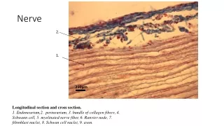
What are myelin sheaths? • Protein-lipid filled cytoplasm of Schwann cells • Neurilemma: outermost part w/nucleus and cytoplasm • Myelin sheath: inner layers of PM • Protects/insulates axon(never dendrites) • Allow for rapid transmission of action potential
What are myelin sheaths? • Nodes of Ranvier: gaps between adjacent Schwann cells • Oligodendrocytes serve same purpose in CNS • White matter: areas of myelinated (primary fiber tracts) • Gray matter: nerve cell bodies (unmyelinated)
Classify by function or structure Structure Multipolar neurons Most common (99%) Three or more processes Many dendrites, some no axon Bipolar neurons Retina, olfactory mucosa Unipolar One process; divides into proximal and distal branches (both are considered axons) What kinds of neurons are there?
What kinds of neurons are there? • Function • Sensory (afferent) neurons • Conduct toward CNS from skin, internal organs • Usually unipolar; soma located outside CNS • More on sensory receptors in special senses lecture • Motor (efferent) neurons • Conduct away from CNS; multipolar • Cell bodies in CNS • Interneurons (association neurons) • Between sensory and motor neurons; multipolar • Usually entirely in CNS; 99% of all your neurons
What does it mean when a neuron “fires”? • Firing = excitability = action potential = nerve impulse • Recall resting potential of all cells • High K+ in; high Na+ out • Cell is polarized • Cell overall neg. charge inside due to molecules like proteins, RNA, DNA • Charge measured in millivolts • Potential = difference in charge across PM • Current = flow of charge (ions) from one point to another
What lets ions move across the PM? • Membrane ion channels (proteins) • Passive (leakage): always open • Active (gated): usually either opened or closed depending on type of gate • Chemically-gated: ligand-gated • E.g. ACh ion gate • Voltage-gated: open/close in response to change in potential
What causes resting potential in the first place? • Membrane permeability • K+ permeable, but not Na+ permeable • Creates membrane potential • K+ leave cell but Na+ can’t enter • Result: overall neg. charge inside cell • Na+/K+ pump maintains but does not create resting potential • Always a lot of K+ leaking out and a little Na+ leaking in
What is depolarization? • Reduction in membrane potential • Less difference between in- and outside of cell • i.e cells becomes less negative (-70 mV to -50 mV) • Cell can also temporarily become positive • Excitatory event • Hyperpolarization • Cell becomes more negative than normal • e.g. -70 mV to -90 mV • Inhibitory event
What are local potentials? • Short-lived, local changes in membrane potential • Can depolarize or hyperpolarize cell • Ligand-regulated • Graded = magnitude varies w/strength of stimulus • Stronger stimulus = greater voltage change, longer travel of current • Caused when ion gates open due to stimulus
What happens during an action potential? • Follow on graph • Sodium ions arrive at axon hillock • Depolarizes membrane • Threshold reached (-55 mV)
What happens during an action potential? • Voltage-regulated Na+ (fast) gates open • Slow voltage-regulated K+ gates also open • Depolarization begins • Propagation of signal
What happens during an action potential? 5. Na+ gates close (inactivate) above 0 mV - voltage peaks around 35 mV - fully depolarized 6. At voltage peak, K+ gates are finally fully open - repolarization begins at K+ flows out • How is this different from resting potential?
What happens during an action potential? 7. K+ gates closer more slowly than Na+ gates - result: more K+ out than Na+ in - overshoot = hyperpolarization http://www.blackwellpublishing.com/matthews/channel.html
What happens after an action potential? • Refractory period: few millisecs • Time during which can’t stimulate neuron a second time • Happens until recovery of resting potential • Two stages • Absolute refractory period • No new action potential possible • Relative refractory period • Can trigger new action potential if stimulus is very strong
How do action potentials travel down the axon? • Nerve signal = traveling wave of excitation produced by action potentials • Unmyelinated sheaths • Slower transmission • Action potential must open all gates between hillock and synaptic knob • Called continuous conduction
How do action potentials travel down the axon? • Myelinated sheaths • Many times faster transmission • Action potential skips from one node of Ranvier to the next • Called saltatory conduction • http://www.blackwellpublishing.com/matthews/actionp.html
What else influences speed of action potential? • Axon diameter • The larger the diameter, the faster the speed of transmission • Less resistance to current flow with larger diameter Slower transduction Faster transduction
What happens if myelination is lost? • Multiple sclerosis • Autoimmune disease • Usually young adults • Blindness, problems controlling muscles • Ultimately paralysis • Immune system attacks myelin sheaths and nerve fibers • Scar tissue (scleroses) replaces some damaged cells • Other now unmyelinated axons sprout Na+ channels • Accounts for sporadic nature of disease?
What happens when the nerve signal reaches the synaptic knob? • First some terminology • Synapse: junction between two neurons • Use neurotransmitters • Allows for integration/evaluation of information • Presynaptic neuron • Can synapse with next neurons dendrites, soma or axon • Postsynaptic neuron • Synaptic cleft
What are neurotransmitters? • Chemicals which cross synaptic cleft • Communicate with postsynaptic neuron • Over 100 known neurotransmitters • ACh, serotonin, glutamate, aspartate, glycine, GABA, NE, dopamine, histamine • Excitatory or inhibitory
How do other neurotransmitters work? • ACh and some others are ionotropic • Alters membrane potential • Rest are metabotropic • Use secondary messenger (e.g. cyclic AMP) to alter postsynaptic cell metabolism • Neurotransmitter activates cAMP production • For example…
Also: http://www.blackwellpublishing.com/matthews/neurotrans.html
How does a nerve signal stop? • Neurotransmitters usually bind for only about 1 msec • Then detaches, then reattaches, then detaches… • If no new neurotransmitter available, stimulus stops • This can happen one of three ways • Diffusion • Destruction (e.g. AChE) • http://www.microvet.arizona.edu/Courses/MIC420/lecture_notes/clostridia/clostridia_neurotox/movie/botulinum_movie.html • Reuptake • Cocaine • http://www.wnet.org/closetohome/animation/coca-anim2-main.html • SSRIs • http://www.paxil.com/flash/depression.swf
How do neurons integrate multiple signals? • Like a democracy: count the votes! • Mechanisms neurons use to process, store and retrieve information • Postsynaptic potentials • Excitatory postsynaptic potential (EPSP) • Na+ flows in an cancels some of neg. charge • Glutamate, aspartate • Inhibitory postsynaptic potential (IPSP) • Increases neg. charge • Neurotransmitter opens Cl- gates into cell • Glycine, GABA • ACh, NE can be either EPSPs or IPSPs
How do neurons integrate multiple signals? • Summation: adding up postsynaptic potentials • Sum determines if fire or not • Need about 30 EPSPs to reach threshold • Temporal summation: new EPSPs arrive before decay of previous EPSP • Summation exceeds threshold • Spatial summation: several different synapses all emit EPSPs • Enough Na+ enters to reach threshold
What are neuronal circuits? • Pathways among neurons • Diverging circuits • Large scale muscle contraction • Converging circuits • Good for incoming sensory information to converge in one part of brain • Reverberating circuit • Promotes inhalation (when reverberation stops, you exhale) • Parallel after-charge circuit • Seeing light bulb image after closing eyes