COLON
COLON. James Taclin C. Banez, MD. Anatomy / Physiology:. Location, blood supply & venous drainage, lymphatic drainage and nerve supply Function: absorption of fluid and electrolyte Transport and temporary storage of feces. Anatomy / Physiology:. Infectious:. Amebic colitis:

COLON
E N D
Presentation Transcript
COLON James Taclin C. Banez, MD
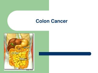
Anatomy / Physiology: • Location, blood supply & venous drainage, lymphatic drainage and nerve supply • Function: • absorption of fluid and electrolyte • Transport and temporary storage of feces
Infectious: • Amebic colitis: Entamoeba histolytica • Primary – colon : secondary – liver • Fecal to oral route: (sexual contact, contaminated water & food) • Abdominal pain, bloody diarrhea, tenesmus, fever Complication: • megacolon / colonic obstruction (partial) ---> AMEBOMA – mass of inflammatory tissue Dx: clin hx / stool exam / indirect hemagglutination test Tx: metronidazole / iodoquinol : rare COLECTOMY
Pseudomembranous colitis: • Complication of antibiotics ---> alteration of normal flora • Overgrowth of Clostridium deficile: • Has cytopathicand enteropathic toxins Develops 6wks after: • Clindamycin • Ampicillin • Cephalosporin Dx: - history - latex fixation test - colonoscopy (Pseudomembrane) Tx: 1. stopped antibiotic ----> metronidazole/vancomycin 2. cholestyramine ---> binds w/ toxin 3. Toxic megacolon---> total colectomy w/ ileostomy
Salmonellosis: Salmonella typhi (typhoid fever) Dx: perforation / bleeding Tx: antibiotic / transfusion / right hemicolectomy w/ or w/o ileostomy • Actinomycosis: A. israeli (gm + anaerobic or microaerophilic bacterium) • Characteristic: - chronic inflammatory induration and sinus formation • Cervicofacial area most frequent site • Abdomen – involves the cecum after AP Tx: surgical drainage and antibiotic (penicillin/ tetracycline)
Volvulus: • Twisting of an air-filled segment of bowel about its narrow mesentery ---> OBSTRUCTION -------> STRANGULATION----> GANGRENE----> PERFORATION ----> PERITONITIS • SIGMOID VOLVULUS (90%): • Redundant sigmoid colon w/ a narrow based mesocolon Sx: colicky abd. pain, distention obstipation, rectal collapse s/sx of dehydration
Volvulus: • SIGMOID VOLVULUS (90%): Dx: FPA – inverted U shaped sausage like loop (diagnostic) • Barium enema – bird beaks deformity • Gangrene – chills/fever, leukocytosis w/ s/x of peritonitis
SIGMOID VOLVULUS (90%): Tx: (-) Signs of Peritonitis: • Reduced the volvulus --->prepare for elective colonic surgery for the recurrence is 40%: - use of flexible scope (+) Signs of Peritonitis / Unsuccessful reduction: • Sigmoidectomy w/ Hartmanns or Divine’s colostomy
Cecal Volvulus: Tx: reduction is impossible --> emergency exploration (+) Gangrene: - right hemicolectomy - end to end ileo-transverse colostomy (-) Gangrene: a) – same – b) Cecopexy c) Pure detorsion (recurrence 7 – 15%) • Transverse colon volvulus: • Rare, due to it’s broad based and short mesentery Tx: resection of redundant transverse colon
DIVERTICULOSIS: Abnormal pouch from the wall of a hollow organ Types: • True diverticula (rare) – right side • False diverticula (common) – due to low fiber diet: left side • Rare before 30y/o; common > 75 y/o • Female > Male Etiology: • Unknown • Theories by Painter et al: • Contraction ring (thickening of circular muscle) • Depletion of dietary fibers ---> narrow lumen • Deteriorating integrity of the bowel wall; elderly has lower tensile strength, lowest in the sigmoid)
Pathology: Site: arteriole penetrates the mesenteric side of the antimesenteric teniae coli: Sigmoid (50%) Descending colon (40%) Entire colon (2-10%) DIVERTICULOSIS:
DIVERTICULOSIS: Clinical Manifestation: • Majority are asymptomatic • Symptomatic patients: • Uncomplicated painful diverticular dse. • (+) LLQ pain and tenderness; • (+) change in bowel habits • (-) rebound tenderness • (-) fever nor leukocytosis Dx: Gastrografin enema Tx: high fiber diet
Complicated diverticular disease: • Diverticulitis / Peridiverticulitis: • Infected diverticula • Diverticula is filled up ---> obstructed ---> mucus secretion and bacteria ---> inflammation at the apex ---> unresolved --> extend intramurally ---> perforate.
Complicated diverticular disease: • Diverticulitis / Peridiverticulitis: Sx: - left lower abd. pain / chills & fever / bowel habit changes - (+) abd. Tenderness, distension if w/ partial obstruction - para-rectal tenderness - frequency / urgency of urination (inflamed bladder)
Complicated diverticular disease: Diverticulitis / Peridiverticulitis: Dx: Cln. Hx. Ct scan of the abd / utrasonography (thickened wall & abscess can be seen) Contrast enema / sigmoidoscopy (risk of spreading infection)
Complicated diverticular disease: • Diverticulitis / Peridiverticulitis: Tx: • NPO or liquid diet • Broad spectrum antibiotic • Meperidine (not morphine) • If improved endoscopy to r/o CA
Complicated diverticular disease: • Perforated Diverticulitis: Sx: - similar to appendicitis (Phlegmon mass) - (+) pneumoperitoneum Classification of perforated diverticulitis (Hinchy) Stage I: abscess confined by mesentery of colon Stage II: pelvic abscess Stage III: generalized peritonitis Stage IV: fecal peritonitis
Complicated diverticular disease: • Perforated Diverticulitis: Tx: initial none operative: - NPO / IVF / Broad spectrum antibiotic/ meperidine Stage I & II: (+) improvement elective Surgery (4 wks) (-) improvement percutaneous drainage (-) improvement ---> Surgery
Complicated diverticular disease: • Perforated Diverticulitis: Stage III & IV: explore after initial resuscitation a. sigmoidectomy w/ primary anastomosis b. sigmoidectomy w/ Hartmann’s colostomy c. resection w/ primary anastomosis w/ proximal diverting stoma
Complicated diverticular disease: • Obstructing diverticulitis: • 90% partial – due to spasm, edema & ileus • 10% complete – fibrosis and stenosis • S/Sx: of large intestinal obstruction • Tx: conservative mx (3-5 days) ---> (-) response -----> cecum dilates to 10-12 cm. ---> surgery.
Complicated diverticular disease: Acute hemorrhage: Due to erosion of the peridiverticular arteriole by inspissated stool w/in the diverticulum and thinning of the tunica media
Clinical Manifestation: Symptomatic patients: Complicated diverticular disease: Acute hemorrhage: Resuscitate the patient Locate the site of bleeding (Tc labeled RBC/selective arteriography) Vasopressin infusion, transcatheter emboli infusion using gelfoam Colonoscopy Tx: segmental resection / blind subtotal colectomy DIVERTICULOSIS:
DIVERTICULOSIS: Clinical Manifestation: • Symptomatic patients: • Complicated diverticular disease: • Fistula formation: • Bladder, vagina, small bowel, skin • Dx: - clin hx & PE (pneumaturia, fecaluria and frequent UTI) - cystoscopy, IE, speculum exam - methylene blue enema - colonoscopy to r/o CA
DIVERTICULOSIS: Clinical Manifestation: • Symptomatic patients: • Complicated diverticular disease: • Fistula formation: • Tx: - bowel rest w/ TPN or elemental diet - Foley catheter (10 days postop) / antibiotic - placement of ureteral catheter prior to celiotomy - sigmoidectomy w/ primary anastomosis - fistulectomy and closure of secondary opening
Hemorrhage from the Colon: • Diverticular disease • Angiodysplasia (Vascular ectasia, AV malformation, Angiectasia)
ANGIODYSPLASIA • Acquired lesion • Proximal colon (cecum) where tension is greatest (Laplace’s law – tension in the wall is highest in the widest circumference) • Rare < 40y/o; common in elderly • Etiology: - chronic intermittent obstruction of submucosal veins due to repeated muscular contraction
Dx: - Nuclear scan / angiography = (vascular tuft and early filling of veins) - colonoscopy = distinct red mucosal patch ANGIODYSPLASIA
Management of Massive Lower GIB • Bleeding distal to the ligament of Treitz: • Diverticular disease • Angiodysplasia • Inflammatory bowel disease • Ischemic colitis • Tumor • Anticoagulant therapy • Gastroduodenal hge -> can present as rectal bleeding • It is more important to identify the location of the BLEEDING POINT than the immediate diagnosis as the cause.
Diagnostic: Nuclear imaging (bleeding scan/scintigraphy) Technetium-Sulfur Colloid Scan Sensitive (0.5ml/min) Autologous labeled RBC scan Stays in the circulation for as long as 24 hrs (monitoring) (1ml/min bleeding) Mesenteric Angiography Done once patient’s condition is stable and hydration is adequate Identify bleeding point ---> 1ml/min Could be therapeutic ---> Vasopressin/emboli Vascular taft (A) Early filling vein (B) Management of Massive Lower GIB
Management of Massive Lower GIB Diagnostic: • Emergent colonoscopy: • Possible w/ use of GOLYTELY • Therapeutic Treatment: • Restore intravascular volume (85% stop spontaneously) • Persistent --> celiotomy (segmental or total colectomy)
Ischemic Colitis • Due to occlusion of major mesenteric vessel • Thrombosis, embolization, iatrogenic ligation) • Elderly: - contraceptive pills - medical problems: a) cardiovascular disease b) DM c) Rheumatoid arthritis • Splenic flexure – most common site in the colon
Ischemic Colitis: Clinical Syndrome Based on: • Extent of vascular occlusion • Duration of occlusion • Efficiency of collateral circulation • Extent of secondary bacterial invasion • Reversible or Transient Ischemic Colitis: • Partial mucosal slough that healed after 2-3 days • Stricturing Ischemic Colitis: • Arterial occlusion ---> hge’ic infarct of mucosa ---> ulcerates ----> bacterial invasion of bowel ---> fibrosis
Ischemic Colitis: Clinical Syndrome Based on: • Gangrenous ischemic Colitis: • Complete arterial occlusion ---> full thickness infarction ---> gangrene ---> perforation ----> PERITONITIS.
Ischemic Colitis: Symptoms: • Depends on the stage of the lesion • Acute mild to moderate generalized or lower abdominal crampy pain ---> HEMATOCHEZIA • Hyperactive bowel sound ---> silent • Abdominal tenderness ---> persist --->r/o peritonitis
Ischemic Colitis: Diagnosis: • Clinical hx & PE • FPA ---> adynamic ileus (stops at the involved segment); Pneumoperitoneum • Contrast enema (water soluble) - thumb printing in the mucosa • Endoscopy (risky)
Ischemic Colitis: Treatment: • Emergency celiotomy - segmental resection w/ primary anastomosis or colostomy
Megacolon: • Large colon due to chronic dilatation, elongation and hypertrophy of the colon • Due to chronic partial colonic obstruction w/ associated chronic constipation • Degree of megacolon is proportional to duration of obstruction
Megacolon: • Congenital Megacolon (Hirschsprung disease) • Congenital absence of ganglion cells in the myenteric plexus (submucosa) of the bowel (aganglionosis) • Usually involves the rectosigmoid • Must be sent to Patho and confirm the presence of ganglion • Acquired megacolon • Chaga’s disease (trypanosoma cruzi) • Neurologic disorders / psychotic patients • Cut higher than 2 cm
Fecal impaction: • Is the arrest and accumulation of the feces in the rectum or colon (dehydrated feces). • Overflow diarrhea w/o relief of the sense of rectal fullness • Result to stercoral ulcer (in the plating) --> bleeding and perforation Mx: - tap water enema / manual extraction - hot sitz bath
Inflammatory Bowel Diseases: • Ulcerative colitis (Mucosal Ulcerative Colitis / Idiopathic Ulcerative Colitis): • involve the colonic mucosa – only the colon • male > female • limited to the colon and rectum • Chronic inflammation of GI tract • Crohn’s Disease (Chronic Interstitial Enteritis/Regional Ilietis): • transmural inflammation anywhere in the GIT – affects entire wall • extraintestinal symptoms proceeds those of intestinal symptoms • female > male • Chronic inflammation of GI tract
Chronic Ulcerative Colitis: Mild & Mod. acute findings: mucosal edema crypt abscess rectal involvement Severe acute disease: Pseudopolyps w/ marked mucosal inflammation & edema Late changes: Discrete ulcers, pus Inflammatory Bowel Diseases:
Crohn’s Disease: Early findings: rectal sparing perianal disease aphthous ulceration Moderate changes: linear ulcers cobblestoning skip lesions Late changes: Contact bleeding Confluent ulcers Strictures & mucosal bridging Inflammatory Bowel Diseases:
Inflammatory Bowel Diseases: Morphologic Features of Crohn’s Disease: Suggestive of Crohn’s Disease: • Focal inflammation in the mucosa • Ileal involvement • Linear or fissuring ulcers • Rectal sparing • Right sided predominance Highly suggestive of Crohn’s disease: • Discontinuous segmental involvement • Aphthoid ulcers Pathognomonic of Crohn’s disease: • Sarcoid granulomas • Transmural inflammation w/ lymphoid nodules • Fistulas (at sites other than anus)
Bowel Involvement in Crohn’s Disease(exam question) • Ileocolic 44% • Colonic 28% • Small bowel only 27% • Anorectal 3%
Inflammatory Bowel Diseases: Extra-intestinal Nonhepatic Manifestations of Idiopathic Inflammatory Bowel Disease: (hypothetical autoimmune disease) (don’t need to memorize this list) Musculoskeletal: Blood & Vascular System • ankylosing spondylitis and sacroiliitis - anemia • peripheral arthritis - thrombocytosis • pelvic osteomyelitis - leucocytosis Skin and Mouth:- hypercoagulable state • erythema nodosum • pyoderma gangrenosum Kidneys & Genitourinary • aphthous stomatitis - nephrolithiasis Eye: - obstructive uropathy • uveitis (iritis) - fistulas to genitourinary • episcleritis Other: - Pleurocarditis & Bronchopulmonary vaxculitis
Medical Therapy for Ulcerative Colitis & Crohn’s Disease • Sulfasalazine – lowers the inflammation • Metronidazole (as well as 2nd gen cephalosporin) • Crohn’s ileocolitis & colitis • Perineal colitis • Not effective in active ulcerative colitis • Corticosteroid – lowers antibody • Oral for mild to moderate active ulcerative colitis and Crohn’s disease • Parenteral for severe or toxic ulcerative colitis or Crohn’s disease • Immunosuppressive agents: • Steroid sparing • Refractory disease