Download

1 / 63
770 likes | 1.22k Vues
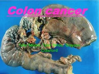
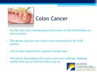
Colon cancer. Epidemiology. 3 rd most common cancer in males and females Accounts for 11% of cancer deaths. In 2000, 130,200 cases (colon and rectum). Lifetime risk 6%. Epidemiology. Rare before the age of 40y, rapid increase at 50y.

E N D
Epidemiology 3rd most common cancer in males and females Accounts for 11% of cancer deaths. In 2000, 130,200 cases (colon and rectum). Lifetime risk 6%.
Epidemiology • Rare before the age of 40y, rapid increase at 50y. • At presentation 37% localized, 37% regional, 20% metastatic. • 1 and 5y survival is 80% and 61% overall. • IBD, FAP, HNPCC, are at inc risk
ascending colon 11% transverse colon 4% descending colon 9% sigmoid colon and rectum 76%
World 50-60% 25-30% Poland 5year survival
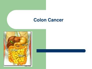
Introduction: Many colorectal cancers are thought to arise from adenomatouspolyps in the colon. These mushroom-like growths are usually benign, but some may develop into cancer over time. Polyps may be small and produce few, if any, symptoms. Regular screening tests can help prevent colon cancer by identifying polyps before they become cancerous.
Your best chance for surviving colorectal cancer is detecting it early. When found early, there is nearly a 90 percent chance for cure.
Symptoms: There often are no symptoms of colorectal cancer in its early stages. Most colorectal cancers begin as a polyp. As polyps grow, they can bleed or obstruct the intestine. When the disease spreads, it is still called colorectal cancer
Symptoms: • rectal bleeding • blood in the stool or toilet after a bowel movement • prolonged diarrhea or constipation • a change in the size or shape of the stool • A change in bowel movement pattern that continues over time • General discomfort in the abdomen (frequent gas pains, cramping pain, feeling of bloating or fullness) • Vomiting • Constant fatigue • Chronic constipation
Risk Factors: • Age: Colorectal cancer is most common in people over 50. • Family history: Your risk is higher with a family history (especially parent, sibling) of colorectal cancer, or adenomatous polyps. • Personal history: Your risk is higher with a personal history of inflammatory bowel disease (Crohn’s disease or colitis), colon cancer, or adenomatous polyps. • Weight: Lack of physical activity and obesity are risk factors.
Diet: A high-fat diet, particularly animal fats, may Increase your risk. Diets high in fruits and Vegetables are thought to decrease your risk. diets high in red and processed meat, as well as those low in fiber, are associated with an increased risk of colorectal cancer. Individuals who frequently eat fish showed a decreased risk • Cigarette smoking and alcohol: Your risk may be higher if you smoke or drink • Physical inactivity: People who are physically active are at lower risk of developing colorectal cancer.
Risk Factors Polyps-Most cancers arise from them. Classified as neoplastic (adenomatous)which are benign or malignant, and nonneoplastic (hyperplastic, mucosal, inflammatory, hamartomaous). Adenomatous polyps found in 33% of people by age 50, 50% by age 70. Most lesions <1cm, 60% single, 40% multiple. Invasive cancer will develop in 24% when untreated.
Polyps Three variants: Tubular(75-87%), tubulovillous (8-15%), Villous(5-10%). Tubulovillous, villous(most in rectum) have most increased risk of cancer 20% and 40% respectively. Size, degree of dysplasia (46% cancer >2cm, 34% in severe dysplasia).
Treatment Endoscopic removal, surveillance every three years. Biopsy if it can’t be removed. Surgery for those not amenable to safe polypectomy (large sessile villous lesions).
Treatment Fungation, ulceration, distortion are contraindications for polypectomy. Colectomy indicated for residual carcinoma, those at high risk for +LN despite complete polypectomy. +margin, poor diff, level 4, vascular, lymphatic invasion. Sessile polyp with invasive cancer should be considered for resection even if no high risk pathologic features. Weigh all against pts medical condition of course.
Hereditary Polyposis Syndromes All have this in common: Multiple intestinal polyps, extraintestinal manifestations. FAP: 1-2% of colon cancer patients. A point mutation of APC gene on chromosome 5, band q21. Polyps found throughout the GI tract but most in colon. Symptoms manifest by ages 16-50. Cancer will develop in all by age 50.
Familial Adenomatous Polyposis (FAP) Familial adenomatous polyposis (FAP) is a genetic condition where affected individuals will develop hundreds to thousands of polyps If a parent has FAP, each child has a 50% (or, 1 in 2) chance of inheriting FAP. Each child also has a 50% chance of not inheriting FAP. FAP does not skip generations. Both males and females are equally likely to be affected. Therefore, if you have FAP, your children each have a 1 in 2 chance of having FAP.
Hereditary Polyposis Syndromes Gardner’s Syndrome: Variant of FAP. Colonic and extracolonic manifestations. Periampulary lesions, duodenal lesions, gastric polyps. Ocular, cutaneous, skeletal (retinal, mandible, jaw, teeth, sebaceous cysts). Desmoids, hepatoblastoma, thyroid cancer, Turcot’s syndrome (brain).
Hereditary Nonpolyposis Syndromes Lynch I and II. Occurs five times more frequently than familial polyposis. 1-5 % of colon cancers. Lynch I just colon, Lynch II also involves endometrium, ovary, stomach, small bowel, biliary, pancreas, ureter, renal pelvis. 85% lifetime risk of colon cancer, more right sided cancers (60-70%), earlier (45y), lower stage, better survival, but 20% risk of metachronous, synchronous lesions.
Inflammatory Bowel Disease Ulcerative colitis carries a risk of colorectal carcinoma 30 times greater than general population. Risk increases with duration of disease. After 30 years, risk increases to 35% Crohn’s disease associated with 10-20 fold increased risk of cancer. Need to do surveillance in these population.
Previous Colon Cancer A second primary colon cancer is three times more likely to develop in patients with a history of colon cancer. Metachronous lesions develop in 5-8% of patients.
History of First-Degree Relatives People with first-degree relatives with colorectal cancer have a 1.8-8 fold increase risk of colorectal cancer. Risk is higher if more than one relative affected. Risk is higher if developed in the relative at a young age.
Pathology >90% adenocarcinomas. Four morphologic variants. Ulcerative (most common), exophytic (polypoid, fungating), annular (classic applecore), submucosal infiltrative(linnitus type). Grading system 1-3. Most developed to least differentiated glandular structures.
Staging A- to submucosa only B1- to muscularis only B2- thru wall, not adjacent. B3- Adjacent organs involved. C1- B1 plus LN C2- B2 plus LN C3- B3 plus LN D- Distant mets
A -- 95 - 100 % • B -- 72 - 80 % • C -- 26 - 34 % • D -- 0 - 2 %
Staging-TNM T1 invades submucosa T2 invades muscularis T3 invades subserosa T4 invades organs outside N1- 1-3 nodes N2- 4 or more nodes N3- central nodes M0- no mets M1- distant mets
Clinical Presentation Bleeding, pain, bowel habit changes, weight loss, anorexia, nausea, vomiting, fatigue, anemia. Right upper quadrant pain, fevers sweats, hepatomegaly, ascites, effusions, adenopathy(METS). Obstruction(5-15%) increases risk of death 1.4 fold. Perforation (6-8%) increases it 3.4 fold. Stage I 15%, Stage II 30%, Stage III 20%, Stage IV 25%. Obstruction less common on right side.
DIAGNOSIS: Colorectal cancer screening rates remain low. Therefore, screening for the disease is recommended in individuals who are at increased risk. There are several different tests available for this purpose.
Continue • Digital rectal exam (DRE): The doctor inserts a lubricated, gloved finger into the rectum to feel for abnormal areas. It only detects tumors large enough to be felt in the distal part of the rectum but is useful as an initial screening test. • Fecal occult blood test (FOBT): a test for blood in the stool. Two types of tests can be used for detecting occult blood in stools i.e. guaiac based (chemical test) and immunochemical. The sensitivity of immunochemical testing is superior to that of chemical testing without an unacceptable reduction in specifity.
Endoscope: • Sigmoidoscopy: A lighted probe (sigmoidoscope) is inserted into the rectum and lower colon to check for polyps and other abnormalities. • Colonoscopy: A lighted probe called a colonoscope is inserted into the rectum and the entire colon to look for polyps and other abnormalities that may be caused by cancer. A colonoscopy has the advantage that if polyps are found during the procedure they can be immediately removed. Tissue can also be taken for biopsy.
Diagnosis Scope, Chest X-ray, Complete blood count, CEA, Localized Fibrous Tumors Preop CT scan? Some get it for abnormal LFTs only (but only 15% of liver mets have abnormal LFTs). Others will get it if large bulky tumors to see about adjacent organs, LN. 10% of mets are missed with preoperative and operative evaluations, IOUS best for this.
Diagnosis 15-20% liver mets not palpable. Preop CEA reflects prognosis, disease extent (over 10-20 poor) CEA may not be elevated in poorly differentiated or rectal cancers. CEA really only good for follow up.
Rectal Cancer In addition to History&Physical, CXR, CBC, LFTs, EUS, Proctoscopic exam, full colonoscopy, CT scan should be done for rectal cancer. Accurate preoperative staging critical because stage may influence treatment decisions such as trans anal excision, preop chemoradiation.
Rectal Cancer EUS is most accurate tool in determining tumor stage with all layers identified with 67-93% accuracy. Differentiating T1 from T3 easy but T2 from T3 harder. Limitations of EUS: operator experience, differentiating LN vs.blood vessels, post radiation changes, stenotic lesions, overstaging (10-15%), understaging (1-2%). Superior to CT or MRI for depth of tumor.
Rectal Cancer • Lymph node staging more difficult. EUS 62-83% accurate, CT scan 35-73% accurate. • All these tests pick up size of LN only. • 50-75% of involved LN are normal in size, so may not be picked up. Similarly, enlarged LN may be inflammatory, so false negative. • LN> 3mm and hypoechoic are likely to have malignancy, also FNA might help under EUS guidance.
Rectal Cancer CT scanning of abdomen and pelvis is important for other organ involvement, and distant spread. CT is better than EUS for contiguous organ involvement.
Pathology: The pathology of the tumor is usually reported from the analysis of tissue taken from a biopsy or surgery. A pathology report will usually contain a description of cell type and grade. The most common colon cancer cell type is adenocarcinoma which accounts for 95% of cases. Other, rarer types include lymphoma and squamous cell carcinoma.
Cancers on the right side (ascending colon and cecum) tend to be exophytic, that is, the tumour grows outwards from one location in the bowel wall. This very rarely causes obstruction of feces, and presents with symptoms such as anemia. Left-sided tumours tend to be circumferential, and can obstruct the bowel much like a napkin ring.