Download
1 / 49
490 likes | 574 Vues
Explore the intricate processes of glycogen metabolism, including synthesis and degradation pathways in the well-fed state. Learn how glucose is stored as glycogen in liver and muscles, and how it is mobilized for energy. Discover the pivotal role of enzymes and hormones in regulating glycogen levels for optimal bodily function.

E N D
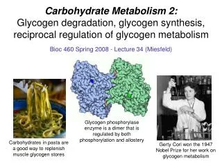
METABOLISM OF CARBOHYDRATES: SYNTHESIS AND DEGRADATION OF GLYCOGEN
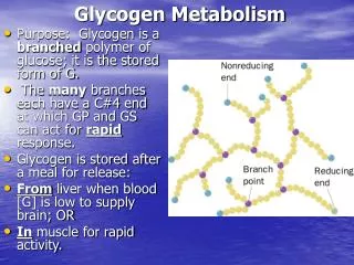
GLYCOGEN SYNTHESIS AND DEGRADATION In the well-fed state the glucose after absorption is taken by liver and deposited as a glycogen Glycogen is a very large, branched polymer of glucose residues that can be broken down to yields glucose molecules when energy is needed Most glucose residues in glycogen are linked by a-1,4-glyco-sidic bonds, branches are created by a-1,6-glycosidic bonds
Glycogen serves as a buffer to maintain blood-glucose level. Stable blood glucose level is especially important forbrain where it is the only fuel. The glucose from glycogen is readily mobilized and is therefore a good source of energy for sudden, strenuous activity. Liver (10 % of weight) and skeletal muscles (2 %) – two major sites of glycogen storage Glycogen is stored in cytosolic granules in muscle and liver cells of vertebrates
Glucose-6-phosphate is the central metabolite in the synthesis and decomposition of glycogen. In the well-fed state glucose is converted to glucose-6-phosphate, which is the precursor for the glycogen synthesis. The glucose-6-phosphate derived from the breakdown of glycogen has three fates: (1) glycolysis; (2) pentose-phosphate pathway; (3) convertion to free glucose for transport to another organs.
DEGRADATION OF GLYCOGEN Glycogenolysis- degradation of glycogen The reaction to release glucose from polysaccharide is not simple hydrolysis as with dietary polysaccharides but cleavage by inorganic phosphate – phosphorolytic cleavage Phosphorolytic cleavage or phosphorolysisis catalyzed by enzyme glycogen phosphorylase There are two ends on the molecules of starch or glycogen: a nonreducing end (the end glucose has free hydroxyl group on C4) and a reducing end (the end glucose has an anomeric carbon center (free hydroxyl group on C1)
Glycogen phosphorylase removes glucose residues from the nonreducing ends of glycogen Acts only on a-1-4 linkages of glycogen polymer Product is a-D-glucose 1-phosphate (G1P) Cleavage of a glucose residue from the nonreducing end of glycogen
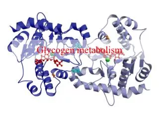
Structure of glycogen phosphorylase (GP) • GP is a dimer of identical subunits (97kD each) • Catalyticsites are in clefts between the two domains of each subunit • Bindingsites for glycogen, allosteric effectors and a phosphorylation site • Two forms of GP • Phosphorylase a (phospho- rylated) active form • Phosphorylase b (dephospho- rylated) less active
GP catalyzes the sequentialremoval of glucose residues from the nonreducing ends of glycogen • GP stops 4 residues from an a 1-6 branch point • Tranferase shifts a block of three residues from one outer branch to the other • A glycogen-debranchingenzyme or 1,6-glucosidase hydrolyzes the 1-6-glycosidic bond • The products are a free glucose-1-phosphate molecule and an elongated unbranched chain
Metabolism of Glucose 1-Phosphate (G1P) • Phosphoglucomutasecatalyzes the conversion of G1P to glucose 6-phosphate (G6P)
Glycogen Synthesis • Synthesis and degradation of glycogen require separate enzymatic steps • Cellular glucose converted to G6P by hexokinase • Three separate enzymatic steps are required to incorporate one G6P into glycogen • Glycogen synthase is the major regulatory step
Glucose 1-Phosphate formation • Phosphoglucomutasecatalyzes the conversion of glucose 6-phosphate (G6P) to glucose 1-phosphate (G1P).
UDP-glucose is activated form of glucose. UDP-glucose is synthesized from glucose-1-phosphate and uridine triphosphate (UTP) in a reaction catalized by UDP-glucose pyrophosphorylase
Glycogen synthase adds glucose to the nonreducing end of glycogen
A branching enzyme forms -1,6-linkages Glycogen synthase catalyzes only -1,4-linkages. The branching enzyme is required to form -1,6-linkages. Branching is important because it increases the solubility of glycogen. Branching creates a large number of terminal residues, the sites of action of glycogen phosphorylase and synthase.
Regulation of Glycogen Metabolism • Muscleglycogen is fuel for muscle contraction • Liverglycogen is mostly converted to glucose for bloodstream transport to other tissues • Both mobilization and synthesis of glycogen are regulated by hormones • Insulin, glucagon and epinephrine regulate mammalian glycogen metabolism
Hormones Regulate Glycogen Metabolism Insulin • Insulin isproduced by b-cells of the pancreas (high levels are associated with the fedstate) • Insulin increases rate of glucose transport into muscle, adipose tissue via GluT4 transporter • Insulin stimulates glycogensynthesis in the liver via the second messenger phosphatidylinositol 3,4,5-triphosphate (PIP3)
Glucagon • Secreted by the a cells of the pancreas in response to lowbloodglucose (elevated glucagon is associated with the fastedstate) • Stimulates glycogendegradation to restore blood glucose to steady-state levels • Only liver cells are rich in glucagon receptors and therefore respond to this hormone
Epinephrine (Adrenalin) • Released from the adrenal glands in response to sudden energy requirement (“fight or flight”) • Stimulates the breakdownofglycogen to G1P (which is converted to G6P) • Increased G6P levels increase both the rate of glycolysis in muscle and glucoserelease to the bloodstream from the liver and muscles • Both liver and muscle cells have receptors to epinephrine
Reciprocal Regulation of GlycogenPhosphorylase and Glycogen Synthase • Glycogen phosphorylase (GP) and glycogen synthase (GS) control glycogen metabolism in liver and muscle cells • GP and GS are reciprocallyregulated both covalently and allosterically (when one is active the other is inactive) • Covalent regulation by phosphorylation (-P) and dephosphorylation (-OH) • Allosteric regulation by glucose-6-phosphate (G6P)
Reciprocal Regulation of GP and GS COVALENTREGULATION Activeform “a” Inactiveform “b” Glycogen phosphorylase -P -OH Glycogen synthase -OH -P ALLOSTERICREGULATION by G6P GP a (active form) - inhibited by G6P GS b (inactive form) - activated by G6P
Activation of GP and inactivation of GS by Epinephrine and Glucagone
Gluconeo- genesis Carbohydrates provide a significant portion of human caloric intake
All cells are dependent on glucose. Glucose level in blood plasma must be stable. Brain is especially sensitive to the decrease of glucose level (the daily glucose requirement of the brain in a typical adult human being is about 120 g). Red blood cells use only glucose as a fuel. 160 g of glucose needed daily by the whole body. The amount of glucose present in body fluids is about 20 g, and that readily available from glycogen is approximately 190 g. During period of fasting glycogen in liver is mobilized but it only lasts 12 to 24 hours and this source of glucose may not fulfill metabolic need. During a longer period of starvation organism must synthesize glucose from smaller noncarbohydrate precursor molecules.
Gluconeogenesis–synthesis of glucose from noncarbohydrate precursors • Liver and kidney are major sites of glucose synthesis • Main precursors: lactate, pyruvate, glycerol and some amino acids • Under fasting conditions, gluconeogenesis supplies almost all of the body’s glucose • Gluconeogenesis – universal pathway. It present in animals, microorganisms, plants and fungi • Plants synthesize glucose from CO2 using the energy of sun, microorganisms – from acetate and propionate
Gluconeogenesis is not a Reversal Glycolysis In glycolysis, glucose is converted into pyruvate; in gluconeogenesis, pyruvate is converted into glucose. However, gluconeogenesis is not a reversal of glycolysis. There are three irreversible reactions in glycolysis catalyzed by hexokinase, phosphofructokinase, and pyruvatekinase. 1. Glucose + ATP glucose-6-phosphate + ADP(hexokinase) G = -8 kcal mol-1 3. Fructose-6-phosphate + ATP fructose-1,6-biphosphate + ADP(phosphofructokinase) G = -5.3 kcal mol-1 10. Phosphoenolpyruvate + ADP pyruvate + ATP(pyruvate kinase) G = -4 kcal mol-1 These three reactions must be bypassed in gluconeogenesis
Bypassed Reactions in Gluconeogenesis 1. Phosphoenolpyruvate is formed from pyruvate by way of oxaloacetatethrough the action of pyruvatecarboxylase and phosphoenolpyruvatecarboxykinase. Pyruvate + CO2 + ATP + H2O oxaloacetate + ADP + Pi + 2H+ Oxaloacetate + GTP phosphoenolpyruvate + GDP + CO2 2. Fructose 6-phosphate is formed from fructose 1,6-bisphosphate. Enzyme - fructose 1,6-bisphosphatase. Fructose 1,6-bisphosphate + H2O fructose 6-phosphate + Pi 3. Glucose is formed by hydrolysis of glucose 6-phosphatein a reaction catalyzed by glucose 6-phosphatase. Glucose 6-phosphate + H2O glucose + Pi
Gluconeogenesis The distinctive reactions are shown in red.
Bypass I: Pyruvate Phosphoenolpyruvate The first step in gluconeogenesis is the carboxylation of pyruvate to form oxaloacetate at the expense of a molecule of ATP. Enzyme pyruvatecarboxylaseis present only in mitochondria. Pyruvate is transported into mitochondria from cytoplasm; thepart of pyruvate is formed in mitochondriafrom amino acids. Essential cofactor of pyruvate carboxylase is biotin, which serves as a carrier of CO2. Biotin-binding domain of pyruvate carboxylase Structure of carboxybiotin
Pyruvate carboxylase is allosterically activated by acetyl CoA. Accumulation of acetyl CoA from fatty acid oxidation signals abundant energy, and directs pyruvate to oxaloacetate for gluconeogenesis. Pyruvate carboxylase reaction biotin This reaction takes place in mitochondria matrix.
Oxaloacetate is polar molecule and can not pass through the mitochondria membrane into cytoplasm Therefore it is reduced: oxaloacetate + NADH2 malate + NAD+Enzyme – malate dehydrogenase Malate passes through the mitochondria membrane into cytoplasm and again oxidized to oxaloacetate (enzyme malate dehydrogenase): malate + NAD+ oxaloacetate + NADH2 Cytoplasmic oxaloacetate is decarboxylated to phosphoenolpyruvate by phosphoenolpyruvate carboxykinase
Phosphoenolpyruvate carboxykinase reaction Reaction takes place in the cytosol. In decarboxylation reaction GTP donates a phosphoryl group. Oxaloacetate is simultaneously decarboxylated and phosphorylated by phosphoenolpyruvate carboxykinase. One molecule of ATP and one molecule of GTP were spent to lift pyruvate to the energy level of phosphoenlpyruvate.
Bypass II: Fructose 1,6-bisphosphate Fructose 6-phosphate • A metabolically irreversible reaction • The enzyme responsible for this step is fructose 1,6-bisphosphatase • F1,6BPase is allosterically inhibited by AMP and fructose 2,6-bisphosphate (F2,6BP)
Bypass III: Glucose 6-phosphate glucose • In most tissues, gluconeogenesis ends with the formation of glucose 6-phosphate (G-6P). • Glucose 6-phosphate, unlike free glucose, cannot diffuse out of the cell. • The generation of free glucose is controlled in two ways: • enzyme responsible for the conversion of glucose 6-phos-phate into glucose, glucose 6-phosphatase,is regulated; • enzyme is present only in tissues whose metabolic duty is to maintain blood-glucose homeostasis — liver and to a lesser extent kidney, pancreas, small intestine.
The final step in the generation of glucose does not take place in the cytosol. G-6-P is transported into the lumen of the endoplasmic reticulum, where it is hydrolyzed by glucose 6-phosphatase, which is bound to the membrane. Glucose 6-phosphatasereaction
Ca2+-binding stabilizing protein is essential for phosphatase activity. Glucose and Pi are then shuttled back to the cytosol by transporters. Generation of glucose from glucose 6-phosphate. Several proteins play a role in the generation of glucose. T1 transports G-6-P into the lumen of the ER; T2 and T3 transport Pi and glucose respectively back into the cytosol. SP – Ca-binding protein.
The Net Reaction of Gluconeogenesis 2 Pyruvate + 2 NADH + 4 ATP + 2 GTP + 6 H2O Glucose + 2 NAD+ + 4 ADP + 2 GDP + 6 Pi + 2H+ G°' = -9 kcal mol-1 Six nucleotide triphosphate molecules are hydrolyzed to synthesize glucose from pyruvate in gluconeogenesis, whereas only two molecules of ATP are generated in glycolysis in the conversion of glucose into pyruvate. The extra cost of gluconeogenesis is four high phosphoryl-transfer potential molecules per molecule of glucose synthesized from pyruvate.
Subcellular Locations of Gluconeogenic Enzymes • Gluconeogenesis enzymes are cytosolic except: • (1) Glucose 6-phosphatase (endoplasmic reticulum) • (2) Pyruvatecarboxylase (mitochondria) • (3) Phosphoenolpyruvatecarboxykinase (cytosol and/or mitochondria)
Regulation of Gluconeogenesis Gluconeogenesis and glycolysis are reciprocally regulated - within a cell one pathway is relatively inactive while the other is highly active. The amounts and activities of the distinctive enzymes of each pathway are controlled. The rate of glycolysis is determined by the concentration of glucose. The rate of gluconeogenesis is determined by the concentrations ofprecursors of glucose.
AMP stimulates phospho-fructokinase, whereas ATP and citrate inhibit it. Fructose 1,6-bisphosphatase is inhibited by AMP and activated by citrate. Fructose 2,6-bisphosphatestrongly stimulatesphospho-fructokinase 1 and inhibits fructose 1,6-bisphosphatase. During starvation, gluconeo-genesis predominates because the level of F-2,6-BP is very low. High levels of ATP and alanine, which signal that the energy charge is high and that building blocks are abundant, inhibit the pyruvate kinase. Pyruvate carboxylase is activated by acetyl CoA and inhibited by ADP. ADP inhibits phosphoenol-pyruvate carboxykinase. Gluconeogenesis is favored when the cell is rich in biosynthetic precursors and ATP.
Regulation of the Enzymes Amount by Hormones Hormones affect gene expression primarily by changing the rate of transcription. Insulin, which rises subsequent to eating, stimulates the expression of phosphofructokinase and pyruvate kinase. Glucagon, which rises during starvation, inhibits the expression of these enzymes and stimulates the production of phosphoenolpyruvate carboxykinase and fructose1,6-bisphosphatase. Transcriptional control in eukaryotes is much slower than allosteric control; it takes hours or days in contrast with seconds to minutes.
Precursors for Gluconeogenesis • Any metabolite that can be converted to pyruvate or oxaloacetate can be a glucose precursor • Major gluconeogenic precursors in mammals: • (1) Lactate • (2) Most aminoacids (especially alanine), • (3) Glycerol (from triacylglycerol hydrolysis)
Lactate • Glycolysis generates large amounts of lactate in active muscle • Red blood cells steadily produce lactate • Lactate produced by active skeletal muscle and erythrocytes is a source of energy for other organs • The plasma membranes of some cells, particularly cells in cardiac muscle, contain carriers that make them highly permeable to lactate and pyruvate. • Lactate and pyruvate diffuse out of active skeletal muscle into the blood and then into these permeable cells. • Once inside these well-oxygenated cells, lactate can be reverted back to pyruvate and metabolized through the citric acid cycle and oxidative phosphorylation to generate ATP. • The use of lactate in place of glucose by these cells makes more circulating glucose available to the active muscle cells. • Excess lactate enters the liver.
The Cori Cycle Liver lactate dehydrogenase converts lactate to pyruvate (a substrate for gluconeogensis) Glucose produced by liver is delivered to peripheral tissues via the bloodstream Contracting skeletal muscle supplies lactate to the liver, which uses it to synthesize glucose. These reactions constitute the Cori cycle
Amino Acids • Carbon skeletons of most amino acids are catabolized to pyruvate or citric acid cycle intermediates • The glucose-alanine cycle:(1) Transamination of pyruvate yields alanine which travels to the liver(2) Transamination of alanine in the liver yields pyruvate for gluconeogenesis(3) Glucose is released to the bloodstream