Download
1 / 1
10 likes | 157 Vues
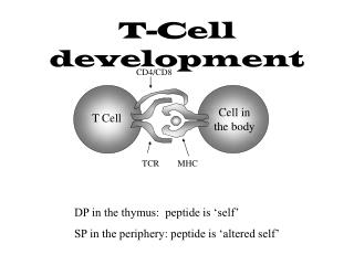
NO cDNA. NO RT. 1000. 900. (Control). (Control). 800. 700. 600. 500. 400. 300. 200. 100. N. DN CELL. N. DP CELL. N. N. CD8 CELL. CD4 CELL. Ex3. Ex5. Ex5. Ex5. Ex4. Ex4. Ex4. Ex3. Ex3. ATG. ATG. ATG. Ex1. Ex1. Ex2. Ex2. Ex2. P. CD4. CD4.

E N D
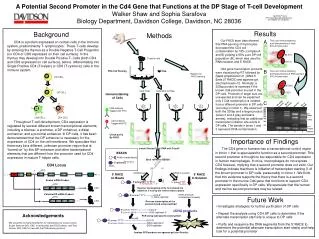
NO cDNA NO RT 1000 900 (Control) (Control) 800 700 600 500 400 300 200 100 N DN CELL N DP CELL N N CD8 CELL CD4 CELL Ex3 Ex5 Ex5 Ex5 Ex4 Ex4 Ex4 Ex3 Ex3 ATG ATG ATG Ex1 Ex1 Ex2 Ex2 Ex2 P CD4 CD4 A Potential Second Promoter in the Cd4 Gene that Functions at the DP Stage of T-cell Development Walker Shaw and Sophia Sarafova Biology Department, Davidson College, Davidson, NC 28036 Results Background Methods • Our FACS stain data showed that PNA panning of thymocytes decreased the CD4 cell contamination by 78% (compare A and B) yielding a 95% pure DP cell population (B), which was used for RNA isolation and 5’ RACE. • Cd4 gene transcription products were visualized by RT followed by Rapid Amplification of cDNA 5’ Ends (5’ RACE) and agarose gel electrophoresis (C). Normally, a 320bp product is expected if the known Cd4 promoter is used in the DP cells. Products of larger size are unexpected and can be explained only if Cd4 transcription is initiated from a different promoter in DP cells (occuring in intron 1). We observed both the 320bp and a larger product (lanes 3 and 4 gray and black arrows), indicating that an additional transcription initiation site exists in DP cells. The bands in lanes 1 and 2 represent DNA contamination. A Two color immunostaining and flowcytometry of fresh B10.A thymocytes • CD4 is a protein expressed on certain cells in the immune system, predominantly T- lymphocytes. These T-cells develop by entering the thymus as a Double Negative T-Cell Progenitor (no CD4 or CD8 expressed on their cell surface). In the thymus they develop into Double Positive T- Cells (both CD4 and CD8 expressed on cell surface), before differentiating into Single Positive CD4 (T-helper) or CD8 (T-cytotoxic) cells in the immune system. • Throughout T-cell development, CD4 expression is regulated by several different known transcriptional elements, including a silencer, a promoter, a DP enhancer, a distal enchancer, and a proximal enhancer. In DP cells, it has been demonstrated that the DP enhancer is necessary for the expression of CD4 on the cell membrane. We speculate that there may be a different, unknown promoter region that is “turned on” by this DP enhancer and other transcriptional elements that are different from the promoter used for CD4 expression in mature T-helper cells. 84.37% Thymus cells 6.91% B PNA Cell Panning Two color immunostaining and flowcytometry of PNA-panned B10.A thymocytes PNA Reserve for FACS staining 95.58% Cells Float Cells Stick to Plate 1.52% Immunostaining of Cells C RT on Beads Liquid RT CD8 antibody Tagged with FITC 500bp 320bp CD4 antibody Tagged with PE Primer dimers 1 2 3 4 Check purity By FACS Importance of Findings • The CD4 gene in humans has a transcriptional control region in intron 1 that is speculated to function as a second promoter. This second promoter is thought to be responsible for CD4 expression in human macrophages. In mice, macrophages do not express CD4 however, implying that a second promoter does not exist. Our findings indicate that there is initiation of transcription starting 3’ of the known promoter in DP cells, presumably in intron 1. We think that this evidence supports the theory that there is a second promoter in the murine Cd4 gene that functions to support CD4 expression specifically in DP cells. We speculate that the human and murine second promoters may be related. Isolate Panned Cells’ RNA with Trizol® BEADS Avidin-coated beads Bind mRNA to BEADS 5’-primer TTTTTTT CD4 Locus biotin ? … … mRNA bound mRNA in solution 5’ RACE on Beads 5’ RACE In Solution TATA Sil DPe Known mRNA Product 5’ primer TTTTTTT-3’ 5’-primerTTTT 3’-AAAAAAA-----5’ 3’AAAA------5’ … Reverse transcription of the first strand; Cs added on 3’ end by the reverse transcriptase. Potential DP mRNA Product (same products, not attached to beads) 5’-primer TTTT------------CCC-3’ 3’-GGG-capfinder Future Work … Reverse transcription of the second strand using capfinder • Investigate strategies for further purification of DP cells • Repeat the analysis using CD4 SP cells to determine if the alternate transcription start site is unique to DP cells • Purify and sequence the DNA segments from the 5’ RACE to determine the potential alternate transcription start site(s) and help look for a potential promoter 5’-primer TTTT------------------CCC-3’ cDNA products -----------------GGG-capfinder-5’ 3’-primer AAAA Acknowledgements PCR using Cd4 specific exon3 primer exon3 5’-primer TTTT--------------------------------CCC-3’ exon3----capfinder • We would like to thank Amy Becton for maintaining our mouse colony, Susan Sharrow (NCI, EIB) for antibodies and FACS advice, and Terry Guinter (NCI, EIB) for help with the PNA panning protocol. -------------------------------GGG-capfinder-5’ 3’-primer AAAA exon3----capfinder GGG-capfinder Analyze PCR products on agarose gel (see Results)