research article
As our journal we prioritize the highest ethical standards in publishing. We strictly adhere to established publication ethics, ensuring that all submissions undergo a thorough peer review process. Our dedicated reviewers, who are experts in their respective fields, assess the quality, originality, and scientific soundness of each manuscript. This rigorous review process contributes to the credibility and reliability of the research published in our journal.<br>https://thebioscan.com/

research article
E N D
Presentation Transcript
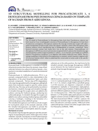
2 (1) : 1-7, 2007 3D STRCUTURAL MODELLING FOR PROCATECHUATE 3, 4 DIOXYGENASEFROMPSEUDOMONASCEPACIABASEDONTEMPLATE OF M CHAIN FROM P . AERUGINOSA D. JAYASREE1, J.VENKATESHWARA RAO1, D. VENKATA KRISHNA RAO1, K. K. KUMAR1, P. B. K. KISHORE3, H. A. NAGARAJARAM2, Y.PRAMEELA DEVI1, M. LAKSHMI NARASU1, 1CentreforBiotechnology,InstituteofScience&Technology,JNTU,Kukatpally-500085,Hyderabad 2Centre for DNA and Finger Printing Diagnostics, Nacharam - Hyderabad 3DepartmentofGenetics,OsmaniaUniversity,Hyderabad-500007 KEY WORDS Multiple sequence alignment Ramachandram Plot Joy alignment Steriochemical equality ABSTRACT The 3D model of Protocatethuate 3,4 dioxygenase beta chain from Pseudomonas cepacia was built, based on X-ray crystallographic structure of Protocatethuate 3,4 dioxygenase M chain from Pseudomonas aeruoginosa. Dioxygenase catalyses the cleavage of molecular oxygen with subse- quent incorporation of both oxygen atoms into organic substrates. Some of the dioxygenase from bacteria catalyse critical ring-opening step in biodegradation of aromatic compounds. These bacterial enzymes contain non- heme ferric iron as the sole cofactor. Protocatethuate 3,4 dioxygenase (3,4PCD) was one of such enzyme recognized which catalyses the intradiol cleavage of protocatechuic acid by oxygen to produce â- carboxy –cis-cis-muconic acid. Studies on 3,4(PCD) found in Pseudomonas aeruginosa is an oligomer with relative molecular mass (587K). The ho- mology modelling is done based on the programmes to annotate protein sequence aligment with 3D structural features which helps to understand the conservation of amino acids in the specific local environment. Received on : 21.10.06 Accepted on : 27.01.07 * Corresponding author substrate are in the ortho position, the ring fission by the intradiol aromatic-ring-cleavage dioxygenases (IARCD) occurs between the two hydroxyl groups (cf. extradiol aromatic-ring-cleavagedioxygenases): INTRODUCTION Protocatechuate3,4-dioxygenaseisamemberofafamily ofbacterialenzymesthatcleavethearomaticringsoftheir substrates between two adjacent hydroxyl groups, a key reactioninmicrobialmetabolismofvariedenvironmental chemicals. Protocatechuate (3, 4-dihydroxybenzene, PCA) is an aromatic compound which is a key intermediate in the degradation of the plant biopolymer lignin and other aromatic compounds. The key step of PCA degradation isthering-cleavageperformedbydioxygenasesaddingboth atoms from molecular oxygen to specific carbon atoms withinthering.Thisstepcanbeperformedbytwodistinct mechanisms; intradiol cleavage and extradiol cleavage. In intradiol cleavage the oxygen atoms are added to the carbons carrying the hydroxyl groups, producing two carboxylategroups.Inextradiolcleavagetheoxygensare addedtoonecarboncarryingahydroxylgroupandanother carrying a hydrogen, resulting in the formation of a carboxylategroupandanaldehydicgroup. The extradiol dioxygenases use Fe(II) to activate oxygen for nucleophilic attack on the aromatic substrate, while the intradiol dioxygenases use Fe(III) to activate the aromatic substrate for an electrophilic attack by oxygen . Mechanismofaction Thearomatic-ring-cleavagedioxygenasesopenthearomatic ringbyincorporatingtwoatomsofdioxygen(O2)intheir substrates,typicallycarryingtwoormorehydroxylgroups on the aromatic ring. If two of the hydroxyl groups of a ARCD enzymes, catechol 1,2-dioxygenase (1,2-CTD; EC 1.13.11.1)andprotocatechuate3,4-dioxygenase(3,4-PCD; EC1.13.11.3),containasingleFeIIIasaprostheticgroup. 1,2-CTD enzymes are oligomers composed of either heterodimers (alphaß)n or homodimers (alphaalpha)n. 3,4-PCD contain equal numbers of alpha and ß subunits andformdifferentquaternarystructuresof(alphaß)n(n= 3 to 12) (Harayama et al., 1992).The sequence similarity between1,2-CTDandthealphaandßsubunitsof3,4-PCD suggestscommonancestryofIARCD(QueandHo,1996). The best-characterised IARCD, 3,4-PCD, catalyses the cleavageofanaromaticringof3,4-dixydroxybenzoateto form a dicarboxylic acid (Que and Ho, 1996).Each of the carboxylategroupscontainsoneoftheoxygenatomsfrom O2 (Harayama and Rekik, 1989). 1
D. JAYASREE et al. The substrate activation mechanism for IARCD is represented in Fig. 1 proteins at the same time. The proteins can be superimposedinordertodeducestructuralalignmentsand compare their active sites or any other relevant parts. The purposes of any molecular modeling programme are tobuild,studyandmanipulate molecules.SYBYLprovides powerfultoolstoaccomplishthesegoals.Thetechniques, both command and menu driven, are simple enough for use on small molecules, yet powerful enough to manipulate large molecules. 5. Structuralvaluation:Procheck&Joyalignment Procheck is a suite of programmes to check the stereochemical quality of protein structures.OY is a program to annotate protein sequence alignments with three-dimensional (3D) structural features. It was developed to display 3D structural information in a sequence alignment and help to understand the conservation of amino acids in their specific local environments. RESULTSANDDISCUSSION 1.TargetSequence :Aproteinsequencewithanaccession numberP15110isselectedastargetsequenceformodelling from P.cepacia with a protein length (a.a) 235 and molecular weight 26550 da. The target sequence is given in Table 1. Table 1: target sequence for modelling from P. cepacia Figure 1: The native enzyme contains a highspin, pentacoordinate FeIIIcentre (I). Upon substrate binding, the solvent -derived ligand and Tyrß147 are displaced by a bidentate catecholate dianion (cf. bidentate catecholate monoanion in extradiol aromatic-ring-cleavage reaction) and the FeIIIcentre (II) remains pentacoordinate. The attack of O2 on the semiquinone radical (III) yields a transient alkylperoxide radical (IV) which combines with FeIIcentre to generate a tridentate alkylperoxo-FeIIIcomplex (V). Decomposition of compound (V) by a Crigeetype rearrangement yields muconic anhydride and a native- like FeIIIcentre (VI). Muconic anhydride is subsequently hydrolysed by an FeIII-bound hydroxide derived from O2(Que and Ho, 1996). MATERIAL AND METHODS Methods and software tools used for modelling: 1. Sequenceretrieval:Pedant(http://pedant.gsf.de/) The pedant genome database provides exhaustive automaticanalysisofgenomicsequencesbyalargevariety of bioinformatics tools. For example the following pre- computed analyses are available to analyse protein function: Blast similarity searches against the non- redundant protein sequence database, motif searches against the Pfam, BLOCKS and PROSITE databases 2. 3DPSSM : (http://www.sbg.bio.ic.ac.uk/~3dpssm/) Fast,web-basedmethodforproteinfoldrecognitionusing 1D and 3D sequence profiles coupled with secondary structure and solvation potential information was done. 3. Multiple sequence alignments & phylogenetic tree Clustal(www.molecularevolution.org/software/clustalx) The Clustal programme was employed as the Clustal programmesarethechoiceforthenovicetomakeadecent phylogeneticanalysisofobtainednucleotideoraminoacid sequences. Clustal can align the sequences and produce output files for drawing trees 4.Modelling:SPDBVIEWER&SYBYL:(http://expasy.org spdbv/) DeepView - Swiss-PdbViewer is an application that providesauserfriendlyinterfaceallowingtoanalyzeseveral TemplateSequence:Thetemplateselectedandusedfor protein model- ling of P15110 3Pcc, is a X-ray crystallized structure Protocatechuate 3,4dioxygenase fromPseudomo- nasputida.3pcc is having 238 residues with a m o l e c u l a r weight of 2662 da and 12 al- pha, 12 beta chains with a resolution of 1.98 A0 . 2. 3D PSSM re- sults for fold recognition of Table 2: 3D PSSM results for flod recognition 2
3D STRCUTURAL MODELLING FOR PROCATECHUATE The target sequence was submitted to fold recognition serverlocatedathttp://bmn.icnet.ac.uk/3dpssm.Fromthe aboveresults(Table2)atemplate3pccwith47%identity of protocatechuate 3, 4 dioxygenase mchain from Pseudomonasputidawitharesolutionof1.98A0,Rvalue of 0.163 and with out any bond breakages in the X-ray crystallography structure satisfying all the properties of a template was found. 3. PDB Blast By using PDB Blast( http:// bioinformatics.ljcrf.edu/pdb blast) very remote homologues structures (Table 3) is detected which helps in constructiong sequence profiles. Sothetargetsequence(P15110)isPDBblastedandresults were found that the target sequence is close structural similarity with 3PCC M chain of 235 a.a length. Table 3: Remote homologues structures using PDB Blast 1. gi|10121001|pdb|1EOA 266 1e-72 34% 176 ChainA, 2. gi|10120999|pdb|1EO9 266 1e-72 34% 176 ChainA, 3. gi|3212804|pdb|3PCC| 207 8e-55 47% 235 Chain M, 4. gi|3212780|pdb|3PCD| 205 4e-54 46% 235 Chain M, 5. gi|10121000|pdb|1EO9 200 9e-53 45% 226 Chain B, 4.Multiplesequencealignment Based on above PDB blast results multiple sequence alignmentwasdonebyusingCLUSTALXprogramme and identifiedtheconservedregions.(Table4)(representedby using : marks). A phylogenetic tree was constructed to trace out the relationships between the sequences. We foundthat3PCGand3PCDaretherootstructureswhich Table 4: Multiple sequence alignment through Clustal X programme 3
D. JAYASREE et al. Figure 2 : Cladogram showing resemblance of structure of 3 PCG and 3 PCD with that of 3PCC 5. Alignment of Target with Template molecule 3DPSSM alignment is given below 4
3D STRCUTURAL MODELLING FOR PROCATECHUATE Figure 5: Ramachandran plot; darkest core regions representing more favourable phi & psi values Figure 6: Verification of sterio-chemical equality by PROCHECK 5
D. JAYASREE et al. have very close resemblance to the template structure (3PCC) (Fig. 2). Based on 3DPSSM, results were found that template is 47%homologoustothetargetandthesecondarystructures like helices, coils and strands are clearly identified in target. So based on the alignment obtained further comparative modelling was done to the target molecule. Model Builder Therefinedsequence–structurealignmentasobtained by 3D-PSSM server was used to construct 3D models of Protocatachuate3,4dioxygenaseusingMODELLER.The structure was refinied and some loop regions were correctedusingmodeller.Thetargetmoleculehasbeen shown before and after the modelling in Figs. 3 & 4. Figure 4 :Target molecule after modelling Loopmodeling Loopsoftenplayanimportantroleindefiningfunctional specificity of a protein frame work. Loop modeling is major factor for determining usefulness of comparative models in application such as Ligand docking. Loop modelling provides the information about core and anchorregions. Target: The anchor regions of the loop which we remodeled, is given in Table 5 Table 5 : The anchor regions of loop and resiedue number Anchorregions Resiedueno 30-45 15 Theregionswhicharetobemodelledare30to45residues were carefully selected in loop database SPDBV was scanned. After modelling loop is subjected to energy Figure 3 :Target molecule before modelling: Figure 7: Structure to structure alignment of the sequence model throught Joy alignment 6
3D STRCUTURAL MODELLING FOR PROCATECHUATE minimization. This stereo chemical quality of the residue inthe remodelled loop as well as those in vicinity was checked by plotting Ramchandran plot (Fig. 5) and after remodelling was again submitted to verify 3D program. Ramchandranplot Itshowsphi-psitorsionanglesforallresidues.The darkest region corresponds to core regions representing more favourablecombinationsofphiandpsivalues.Percentage ofcoreregionsisoneofthebetterguidestostereochemi- cal quality. Compatibilityscoreforthemodelisabovezero.Somodel satisfies the environment.Next, the model was submitted to PROCHECK to verify stereo-chemical quality (Fig. 6). Joyalignment Finally the sequence modelled is submitted to joy server, wherejoyshowsstructuretostructurealignment(Fig.7)., which is given by verifying secondary structure. REFERENCES Harayama, S., Kok, M. and Neidle, E.L. (1992) Functional and evolutionary relationships among diverse oxygenases. Annu. Rev. Microbiol. 46: 565-601. Harayama, S. and Rekik, M. (1989) Bacterial aromatic ring-cleavage enzymes are classified into two different gene families. J. Biol. Chem. 264: 15328-15333. Lippard, S.J. and Berg, J.M. (1994) Principles of Bioinorganic Chemistry. University Science Books, Mill Valley. Nishida, Y., Yoshizawa, K., Takahashi, S. and Watanabe, I. (1992) Reaction mechanism 3,4-dioxygenase. Z. Naturforsch. C 47: 209-214. Ohlendorf, D.H., Orville, A.M. and Lipscomb, J.D. (1994) Structure of protocatechuate 3,4-dioxygenase from Pseudomonas aeruginosa at 2.15 Å resolution. J. Mol. Biol. 244: 586-608. Que, L., Jr. and Ho, R.Y.N. (1996) Dioxygen activation by enzymes with mononuclear non-heme iron active sites. Chem. Rev. 96:2607-2624. of protocatechuate 7
SPECIAL ISSUE OF THE BIOSCAN The Editorial Board of The Bioscan is going to bring about special issue of the journal on 1. Physiology and Endocrinology 2. Ecological Productivity and Energetics Interested Academicians, Researchers and Scientists are requested to contribute to the proposed issue. The pattern of preparation of manuscript will be the same as the instruction to the authors of this journal. 8