Eyes
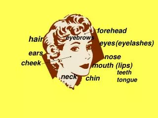
Eyes. External Anatomy. Sensory Organ for vision -Situated in bony, orbital cavity for protection Eyelids= shades that add protection form injury, strong light , dust Eyelashes= hairs to filter dust & dirt. External Anatomy. External Anatomy. Limbus – border b/t the cornea & sclera

Eyes
E N D
Presentation Transcript
External Anatomy • Sensory Organ for vision -Situated in bony, orbital cavity for protection • Eyelids= shades that add protection form injury, strong light , dust • Eyelashes= hairs to filter dust & dirt
External Anatomy External Anatomy
Limbus – border b/t the cornea & sclera • Palpebral fissures – elliptical open space b/t lids • Canthus- corners of the eye where the lids meet, inner & outer • Caruncle – sm. Fleshy mass containing sebaceous glands at inner canthus
Within the upper eyelid • Tarsal plates, connective tissue gives upper lid shape • Meibomian glands, in the plates, lubricate the lids, stops overflow of tears, airtight seal when lids closed
Exposed part of the eye • Conjunctiva, folded envelope b/t eyelids & eyeball • thin mucous membrane, transparent protective covering of the exposed part of the eye. • Palpebral conjunctiva lines the lids, is clear but has sm .bld. Vessels • Bulbar conjunctiva is over eyeball, white sclera show through, merges at limbus with cornea
Lacrimal apparatus – irrigates conjunctiva & cornea • 3 parts • Lacrimal gland, upper, outer corner of eye = tears • Puncta= inner canthus, tear drainage • Nasolacrimal duct= allows tears to drain from puncta to nasolacrimal sac. Tears then empty into the inferior meatus of the nose
Extraoccular muscles • 6 muscles • Attach eyeball to orbit • Straight and rotary movement • Four straight muscles • Superior rectus • Inferior rectus • Lateral rectus • Medial rectus
Two slanting/ oblique muscles • Superior • Inferior Humans have a Binocular, single – image visual system – Eyes normally move as a pair
Eye movement stimulated by Cranial Nerves • III Oculomotor • IV Trochlear • VI Abducens
Internal Anatomy • The eye has 3 layers, the outer & inner layer can be viewed using opthalmascope • Sclera (outer layer) tough, protective, white covering connects with the - • Cornea – transparent, protects pupil & iris – helps focus light on retina
Middle layer • Choroid – dark pigmentation to prevent internal light reflection, supplies bld. to retina • Pupil – PERRLA • Lens – biconvex disc, transparent, thickness controlled by ciliary body, bulges = near; flattens = distant • Anterior chamber – posterior to cornea, anterior to iris & lens, has aqueous humor supplies nutrients & drains wastes
Inner layer – Retina – visual receptive layer – light waves changed to nerve impulses • Retinal structures • Optic disc – retinal fibers meet & form optic nerve, nasal side of retina, creamy yellow orange to pink, round or oval shape, physiologic cup inside the disc for bld.vessels to enter & exit • Retina vessels – paired arteries & veins
Macula – temporal side of fundus, darker pigmented region, surround the fovea centralis • Fovea Centralis- area of sharpest & keenest vision, Very sensitive to light
Visual Pathways & Fields • Objects reflect light • Rays refracted by cornea, aqueous humor, lens, vitreous body and onto retina. • Light stimulus is changed to nerve impulses, travel thru optic nerve to visual cortex in occipital lobe • Image on retina is upside down & reversed. At the optic chiasm retinal fibers cross over. Right side of brain looks at left side of world.
Visual reflexes • Pupillary light reflex – bright light = constriction • Direct light reflex • Consensual light reflex • Fixation – ability to track an object & keep image on the fovea, can be impaired by drugs, alcohol, fatigue & inattention • Accomodation – for near vision = pupil constriction & convergence of eyes
Subjective data • Vision difficulty • Pain • Strabismus, diplopia • Redness, swelling • Watering, discharge • Past history ocular problems • Glaucoma
Glasses/ contacts • Medications • Vision loss- coping mechanisms • Self–care behaviors
Objective data The Physical Exam • Preparation • Position- sitting, head at eye level • Equipment • Snellen eye chart- visual acuity • Handheld visual screener-near vision • Opaque card • Penlight • Applicator stick • Ophthalmoscope
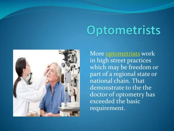
Test visual acuitySnellen eye chart • Stand 20 ft. from chart • Glasses / contacts (Document ) • Remove eye wear, retest • Normal visual acuity is 20/20 – top # is distance person is standing from the chart • Vision 20/30 refer to opthalmologist or optometrist • If unable to see largest letters, move to 10 feet – record as 10/200
Test for near vision • Vision screener • People > 40yrs or difficulty reading • Test each eye with glasses • Hold card 14in. from eyes • Normal result 14 / 14 • Test using any available reading material if no card available
Presbyopia is a normal physiological change in near vision occurs with aging = note if the person moves the card farther away
Test visual fieldsConfrontation test • Compares peripheral vision with a tester who has normal peripheral vision • 2 ft. apart, eye level • Tester & client cover opposite eyes • Tester advances finger in the periphery • Superiorly ( 50 degrees ) • Inferiorly ( 70 degrees ) • Temporally ( 90 degrees )
Inspect Extraoccular Muscle Function • Corneal light reflex • Cover test • Diagnostic positions test • 6 Cardinal Positions of Gaze
Inspect Extraocular Muscle Function • Corneal Light Reflex ( The Hirschberg Test) assesses parallel eye alignment • Shine light toward person’s eyes • Tell to stare directly ahead • Hold light 12 in. away • Light should reflect on both corneas in same spot
Cover Test- detects deviated alignment • Stare straight at examiner’s nose • Cover 1 eye of the person being examined with opaque card • Normally the uncovered eye should maintain a steady, fixed gaze • Covered eye- should stare straight ahead when covered & then uncovered. If muscle weakness exists the covered eye will relax and then jump to fixed position when uncovered..
Diagnostic Positions Test • 6 cardinal positions of gaze – • Determines muscle weakness during movement • Person must hold head steady • Follow movement of object (examiner’s finger, pen etc) only with eyes • Hold object 12 in. from person • Move thru each position, clockwise, hold , then back to center • Normal response= parallel tracking with both eyes
During this test be aware of Nystagmus-fine jerky movement seen around the iris • Mild nystagmus in extreme lateral gaze is normal but not normal in any other position
Inspect External Structures • General – movement & facial expression (squinting?) • Eyebrows – 2(bilateral), symmetrical (look the same; move the same) • Eyelids & Lashes – present, approximate when closed, no redness, swelling, discharge, lesions? • Eyeballs- alignment, ? Protrusion? Sunken? • Conjunctiva & Sclera – moist, glossy, clear, white sclera
Eversion of the upper eyelid FYI – we will not do this examine in lab see pg. 312 for technique – usually done for complaint of eye pain due to foreign body
Lacrimal Apparatus • Person looks down • Using thumbs, slide outer part of upper lid along bony orbit • Note redness or swelling • Press index finger against lacrimal sac at inner canthus • Normal response is slight eversion of lower lid, no tearing or discharge
Anterior Eyeball Structures • Cornea & lens • Iris & pupil • Size & shape • Pupillary light reflex • Accommodation
Cornea & Lens • Shine light from side across cornea • Check smoothness, clarity • Normally no opacities
Iris and Pupil • Iris = flat, round, regular, even color bilaterally. • Pupils = PERRLA • Resting size norm = 3-5mm • 5% population have pupils of 2 diff. Sizes called Anisocoria
Pupillary Light Reflex • Darken room • Person gazes straight ahead • Advance light from the side • Direct light reflex • Consensual light reflex • Measure pupil size before & after light reflex • Measurement R3/1 L3/1 =both pupils measure 3mm in resting state & 1mm with light
Accomodation • focus on distant object -dilatation of pupils • Shift gaze to near object – pupils constrict & converge • Record the normal response to these tests as • PERRLA = Pupils Equal, Round, React to Light and Accomodation