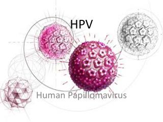
HPV
230 likes | 350 Vues
HPV. Carcinoma of the Cervix. Many risk factors for development of cervical cancer. no routinely used positive predictive biological markers, which identify women at risk of developing high-grade lesions and ultimately invasive cancer. Human Papillomavirus (HPV).

HPV
E N D
Presentation Transcript
Carcinoma of the Cervix • Many risk factors for development of cervical cancer. • no routinely used positive predictive biological markers, which identify women at risk of developing high-grade lesions and ultimately invasive cancer.
Human Papillomavirus (HPV) • Strong association with development of invasive cancer. • >70 types of HPV. • Low risk (6,11). • High risk (16,18,31,33,35,39,45,51,52,56,58,59,66, 68). • Exposure to HPV is followed by a serological response to viral capsid proteins (VLPs). • Immune response is assoc. with persistent HPV infection and is type specific.
E2 E5 L1 E6 L2 E7 E1 E4 8Kbp 0 HUMAN PAPILLOMAVIRUS • small DNA viruses,8kb double stranded genome • a single host may be infected with different HPVs • Two forms of HPV infection of the Cervix • Episomal • Integrated
HPV • Integration of HPV DNA into host loss of E2 orf. • Transcription of E6 and E7 is unregulated. • Transformation events within the cell. • Checkpoint for cell proliferation and transcription is lost.
HPV • Expressed E6 and E7 proteins can then interact with other tumour suppressor genes including p53 and pRB uncontrolled cellular proliferation and malignant transformation. • 3 splice variants of E6 HPV 16 recognised: E6 I, II and III.
E2 E5 L1 E6 L2 E7 E1 E4 Disruption of HPV genome during integration • disruption of E1 to E2 of variable sizes • integration occurs at chromosome ”fragile sites”
Experimental evidence ofHPV transforming capacity RAFT culture experiments with wild type and mutant E6/E7 constructs E6 mutant: in RAFT culture
HPV Cells infected with oncogenic HPV types Immortalisation Uncontrolled cell proliferation
Carcinoma of the cervix • MOLECULAR ONCOLOGY • over 95% of cervical SCCs associated with high risk HPV types (16,18,31,33,45); 40-70% of adenocarcinomas. • HPVs also found in CIN: • 4-6% of normal women HPV 6 and 11 positive. • CIN 1: 10- 30% HPV 6 &11 positive. • CIN 2- 3: 75- 80% HPV 16, 18, 31, 33 positive; 1- 5% HPV 6,11 positive. • HPV E6 and E7 regions can transform epithelial cells and increase cellular levels of cyclins A,B and p34-cdc 2 and cyclin E.
HPV analysis • Who do we screen? • All Women? • HPV as a triage? • How do we screen? • Does HPV analysis give prognostic information? • HPV and other novel biomarkers of disease
Future role for HPV screening • Post introduction of HPV vaccine • vaccines being produced to target HPV 16 and 18 E6/E7 regions. • requirement to monitor HPV status pre and post-vaccination. • possibility of using recombinant • anti-sense PNAs to specifically • target HPV E6 and E6 splice variants.
How do we screen? • HPV analysis • Type • Load • Viral integration
HPV analysis • Technologies available • Hybrid Capture II • PCR generic, (incl. PGYM, GP5 and 6, SYBR green) • Type specific DNA PCR • Solution phase PCR • TaqMan PCR • NASBA (HPV proofer) • In-situ hybridisation (ISH) • Sequence genotyping • In-cell PCR • ICC
HPV analysis • Hybrid Capture II • Liquid based system. • Low and high risk type analysis. • No information in relation to integration. • Indirect load information but NOT quantitative.
Denature NA Hybridise Capture hybrids Label for detection Detect Schematic of Hybrid Capture II
HPV analysis • Hybrid Capture II • Recommended cut-off for the HC-II test is 1 pg viral DNA perml of buffer, equivalent to about 5000 viral genomes. • This cut-off value has been reduced to 0.2 pg/ml but with theintroduction of false positives (Peyton et al). • Data comparing PCR with HC-II found PCR identified HPV in 24.5% of samples, while HC-II detected HPV in13% using the recommended cut-off of 1 pg/ml, and in 22.1%using a cut-off of 0.2 pg/ml.
HPV analysis -PCR • PCR generic / consensus • GP 5 and 6 • PGYM • MY09/11 • SPF10 • GP5 and 6 + SYBR green
Computer-generated amplification plot from a SYBR-green HPV run Detection sensitivity 5-10 copies/reaction
HPV analysis • Type specific PCR • Solution phase PCR • Taq Man q(PCR) • NASBA (HPV proofer) • In-situ hybridisation • HPV genotyping
HPV Beta actin Taq Man PCR Detection sensitivity = 1-2 copies per reaction
HPV analysis • In-situ hybridisation • Cloned HPV subtypes (Zur Hausen) • Automated platforms available. • Commercial probes: • DAKO, Digene, Ventana, etc. Detection sensitivity = 1-5 copies per biopsy