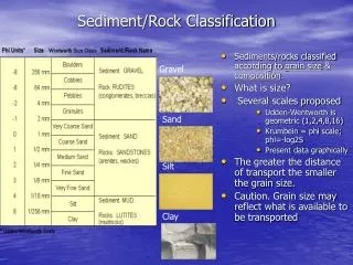
Rock Athroscopy Classification for OCD Lesions - Stability Based Categories
Explore the immobile and mobile categories of OCD lesions with arthroscopic illustrations. Detailed descriptions help in assessing the condition and potential healing.

Rock Athroscopy Classification for OCD Lesions - Stability Based Categories
E N D
Presentation Transcript
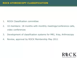
ROCK ATHROSCOPY CLASSIFICATION • ROCK Classification committee • 10 members: 18 months with monthly meetings/conference calls, video conferences • Development of classification systems for MRI, Xray, Arthroscopy • Review, approval by ROCK Membership May 2011
ROCK ATHROSCOPY CLASSIFICATION • GROSS OCD LESION DESCRIPTION • ICRS CARTILAGE CLASSIFICATION • OCD ARTICULAR LESION CONTOUR
ATHROSCOPY CLASSIFICATION: GROSS LESION DESCRIPTION– IMMOBILE and MOBILE • GROSS OCD LESION DESCRIPTION • Focus on appearance of lesion arthroscopically • Lesion stability may have strong relationship to healing potential • 2 Main Categories based upon stability • Immobile (3) • Mobile (5)
GROSS LESION DESCRIPTION– IMMOBILE and MOBILE IMMOBILE Lesions - No observed movement of progeny fragment with respect to the surrounding parent surface upon probing 0. No abnormality detectable arthroscopically (Cue Ball) 1. Cartilage is intact and subtly demarcated (possibly under low light), (Subtle Shadow) 2. Cartilage is demarcated with a fissure, buckle, and/or wrinkle (Wrinkle in the Rug) MOBILE Lesions -- Observed movement of progeny fragment with respect to the surrounding parent surface upon probing 3. Cartilage is intact (Trampoline) 4. Cartilage fissuring at periphery, unable to hinge open (Locked Door) 5. Cartilage fissuring at periphery, able to hinge open (Trap Door) 6. Circumferential fissuring and complete separation, but lesion in situ (Manhole Cover) 7. Exposed sub-chondral bone defect (Crater)
GROSS OCD LESION DESCRIPTION – CUE BALL Illustration Arthroscopic Image Need image 0 - No abnormality detectable arthroscopically IMMOBILE Lesion
GROSS OCD LESION DESCRIPTION – Shadow Illustration Arthroscopic Image Need image Need image 1 - Cartilage is intact and demarcated, but not mobile or ballotable with probing. This may be seen best under low light conditions, or tangential views. IMMOBILE LESION
GROSS OCD LESION DESCRIPTION – Wrinkle in Rug Illustration Arthroscopic Image Need Drawing Need image Need image 2 - Cartilage is demarcated with a fissure, buckle, and/or wrinkle, but not mobile or ballotable with probing (Wrinkle in the Rug) IMMOBILE LESION
GROSS OCD LESION DESCRIPTION – Trampoline Illustration Arthroscopic Image Need image 3 - Cartilage is intact and demarcated, but mobile or ballotable with probing MOBILE LESION
GROSS OCD LESION DESCRIPTION – Locked Door Illustration Arthroscopic Image Need image • 4 - Cartilage fissuring at periphery, but unable to hinge • MOBILE LESION
GROSS OCD LESION DESCRIPTION – Locked Door Illustration Video Image Need image Need image • 4 - Cartilage fissuring at periphery, but unable to hinge • MOBILE LESION
GROSS OCD LESION DESCRIPTION – Trap Door Illustration Arthroscopic Image Need image • 5 - Cartilage fissuring at periphery, with intact hinge (red arrows)
GROSS OCD LESION DESCRIPTION – Locked Door Illustration Video Image Need image Need image • 5 - Cartilage fissuring at periphery, with intact hinge (red arrows) • MOBILE LESION
GROSS OCD LESION DESCRIPTION – Manhole Cover Illustration Arthroscopic Image Need image 6 - Circumferential fissuring, (highlighted by the red arrows) lesion in situ, but can be entirely displaced with a probe. MOBILE LESION
GROSS OCD LESION DESCRIPTION – Crater Illustration Arthroscopic Image Need image Need image 1 - Congruent 2 - Incongruent 3 - Fragmented 4 - Absent 1 - Congruent 2 - Incongruent 3 - Fragmented 4 - Absent 7 - Exposed sub-chondral bone defect Congruent = fragment fits into crater without modifications Incongruent = fragment fits into crater with modifications
GROSS LESION DESCRIPTION– IMMOBILE and MOBILE IMMOBILE Lesions - No observed movement of progeny fragment with respect to the surrounding parent surface upon probing 0. No abnormality detectable arthroscopically (Cue Ball) 1. Cartilage is intact and subtly demarcated (possibly under low light), (Subtle Shadow) 2. Cartilage is demarcated with a fissure, buckle, and/or wrinkle (Wrinkle in the Rug) MOBILE Lesions -- Observed movement of progeny fragment with respect to the surrounding parent surface upon probing 3. Cartilage is intact (Trampoline) 4. Cartilage fissuring at periphery, unable to hinge open (Locked Door) 5. Cartilage fissuring at periphery, able to hinge open (Trap Door) 6. Circumferential fissuring and complete separation, but lesion in situ (Manhole Cover) 7. Exposed sub-chondral bone defect (Crater)
GROSS LESION DESCRIPTION– IMMOBILE and MOBILE IMMOBILE Lesions - No observed movement of progeny fragment with respect to the surrounding parent surface upon probing 0. No abnormality detectable arthroscopically (Cue Ball) 1. Cartilage is intact and subtly demarcated (possibly under low light), (Subtle Shadow) 2. Cartilage is demarcated with a fissure, buckle, and/or wrinkle (Wrinkle in the Rug) MOBILE Lesions -- Observed movement of progeny fragment with respect to the surrounding parent surface upon probing 3. Cartilage is intact (Trampoline) 4. Cartilage fissuring at periphery, unable to hinge open (Locked Door) 5. Cartilage fissuring at periphery, able to hinge open (Trap Door) 6. Circumferential fissuring and complete separation, but lesion in situ (Manhole Cover) 7. Exposed sub-chondral bone defect (Crater) A. Congruent = fragment fits into crater without modifications B. Incongruent = fragment fits into crater with modifications C. Fragmented D. Absent