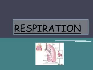
Respiration
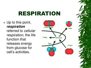
Respiration. Definitions. Breathing(ventilation)- process of moving air into and out of the lungs. Two phases- Inspiration(inhalation) into lungs Expiration(exhalation) out of lungs. Air. Inspired Expired Oxygen 21% 16% Carbon dioxide 0.04% 4%

Respiration
E N D
Presentation Transcript
Definitions • Breathing(ventilation)- process of moving air into and out of the lungs. • Two phases- • Inspiration(inhalation) into lungs • Expiration(exhalation) out of lungs
Air Inspired Expired Oxygen 21% 16% Carbon dioxide 0.04% 4% Nitrogen 78% 78% Water vapour Variable Saturated
12 thoracic vertebrae • 12 pairs of ribs • Sternum or breast bone
Muscles of Respiration • The expansion of the chest during inspiration occurs as a result of muscular activity, partly voluntary and partly involuntary. The main muscles are the Intercostal muscles and the diaphragm. They are assisted by the neck shoulder and abdominal muscles in laboured breathing.
Intercostal Muscles • 11 pairs, found between the ribs. • Arranged in two layers- external and internal. • External- extend in a downwards and forwards direction from the lower border of the rib above to the upper border of the rib below. • Internal- extend in a downwards and backwards direction from the lower border of the rib above to the upper border of the rib below, crossing the external at right angles.
The first rib is fixed, so on contraction all ribs are pulled towards it. Due to their shape they move outwards when pulled upwards. They are stimulated to contract by the Intercostal Nerves.
Muscles of inspiration- diaphragm and external intercostals(elevate ribs and sternum)increase thoracic volume • Muscles of expiration-internal intercostals(depress ribs and sternum)decrease thoracic volume
Diaphragm • A dome-shaped structure which separates the thoracic and abdominal cavities. Supplied be the Phrenic Nerves. Working with the intercostal muscles it ensures that the thoracic cavity enlarges in all directions.
Cycle of Respiration • Occurs about 12-15 times per minute. • Has three phases- inspiration • Expiration • Pause
Inspiration • When the intercostals and the diaphragm contract the capacity of the thoracic cavity increases. This reduces the pressure in the pleural cavity to a lower level than the atmospheric level. The lungs stretch, pressure falls in the alveoli and the air passages. This draws air into the lungs to try to equalise the two pressures. This process is active- requires energy for muscle contraction.
Expiration • Relaxation of intercostals and the diaphragm results in downward and inward movement of the ribs cage and elastic recoil of the lungs. Pressure increases in lungs and exceeds the atmospheric level, therefore air is expelled form the respiratory tract. The lungs retain some air to prevent total collapse .This process is passive- does not require energy. There is now a short pause before the next cycle.
Changing thoracic volume • At the end of a normal ,quiet expiration, the respiratory muscles are relaxed. • During quiet inspiration, contraction of the diaphragm increases the volume of thoracic cavity. This is assisted by contraction of external intercostals, which elevate the ribs and sternum. • Expiration is the result of relaxation of diaphragm and external intercostals with the elastic properties of the lungs causing a decrease in thoracic volume.
During laboured breathing, all of the respiratory muscles are active and contract more forcefully .
PRESSURE CHANGES & AIRFLOW • 2 principles re flow of air in & out of the lungs • Changes in volume result in changes in pressure. The muscles of respiration change thoracic volume and therefore pressure within the thoracic cavity. • Air flows from areas of higher to lower pressure. Air flows through the respiratory passages because of the pressure differences between the outside of the body & the alveoli inside the body.
Lung Recoil • Lung Recoil is the tendency for an expanded lung to decrease in size. 2 reasons for this: • Elastic fibres in the connective tissue of the lungs. • Surface tension of the film of fluid that lines the alveoli which causes the alveoli to recoil & become smaller.
2 FACTORS WHICH PREVENT THE LUNGS FROM COLLAPSING • Surfactant is a mixture of molecules produced by secretory cells of alveolar epithelium. They form a single layer on the surface of the thin fluid layer, reducing surface tension. (Without it the recoil of the alveoli can be 10 x greater). • Pleural pressure. Balloon principle. Balloon expands when pressure inside it is greater than pressure outside. Decreasing pressure in the pleural cavity thus results in expansion of the alveoli.
GAS EXCHANGE • Ventilation is the first stage in respiration. The next stage is the diffusion of gases between the alveoli & the blood in the pulmonary capillaries. • Diffusion across the respiratory membrane is affected by : • Respiratory membrane thickness. • Total surface area of the membrane • Partial pressure of gases across the membrane.
RESPIRATORY MEMBRANE THICKNESS • Thickness of respiratory membrane increases during some respiratory diseases e.g. in pulmonary oedema gases must diffuse through a thicker than normal layer of fluid. Oxygen exchange is affected before carbon dioxide exchange because it diffuses about 20 times less easily.
Surface area • Total surface area of respiratory membrane is about 70 square metres. • Decreased surface area can be a result of surgical removal of lung tissue, or destruction by lung cancer, or degeneration of the alveolar walls by emphysema. Also, collapse of the lung (pneumothorax) drastically reduces the volume of surface area for gas exchange.
PARTIAL PRESSURE • The pressure exerted by a specific gas in e.g. air is called the partial pressure of that gas. Gases diffuse from areas of higher partial pressure towards areas of lower partial pressure until the pressure is equal on both sides.
DIFFUSION OF GASES IN THE LUNGS • Blood returning from tissues into the lungs has a decreased partial oxygen pressure and increased partial carbon dioxide pressure compared with the air in the alveoli. • Oxygen therefore diffuses from the alveoli into the pulmonary capillaries – the blood gains oxygen & loses carbon dioxide. • By the time blood flows through the first third of the pulmonary capillary equilibrium is achieved and diffusion stops.
GAS TRANSPORT IN THE BLOOD • Oxygen Transport – After O2 diffuses across the respiratory membrane into the blood approx. 98.5% of the O2 combines irreversibly with the haemoglobin, becoming oxyhaemoglobin. Approx. 1.5% remains dissolved in the plasma.