Stomach Anatomy
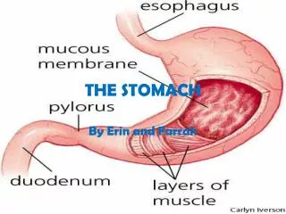
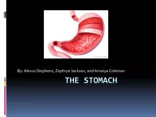
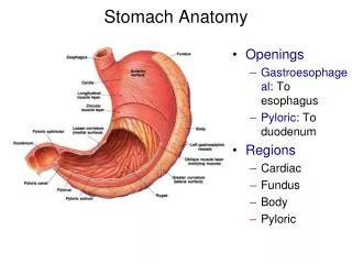
Stomach Anatomy. Openings Gastroesophageal : To esophagus Pyloric : To duodenum Regions Cardiac Fundus Body Pyloric. Stomach Histology:. Layers Serosa or visceral peritoneum: Outermost Muscularis : Three layers Outer longitudinal Middle circular Inner oblique Submucosa Mucosa.

Stomach Anatomy
E N D
Presentation Transcript
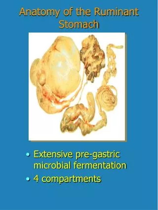
Stomach Anatomy • Openings • Gastroesophageal: To esophagus • Pyloric: To duodenum • Regions • Cardiac • Fundus • Body • Pyloric
Stomach Histology: • Layers • Serosa or visceral peritoneum: Outermost • Muscularis: Three layers • Outer longitudinal • Middle circular • Inner oblique • Submucosa • Mucosa
Stomach Histology • Rugae: Folds in stomach when empty • Gastric pits: Openings for gastric glands • Contain cells
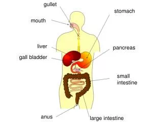
Small Intestine anatomy and Histology • Site of greatest amount of digestion and absorption • Divisions • Duodenum • Jejunum • Ileum: Peyer’s patches or lymph nodules • Modifications • Circular folds or plicaecirculares, villi, microvilli • Cells of mucosa • Absorptive, goblet, granular, endocrine
Small Intestine Secretions • Mucus • Protects against digestive enzymes and stomach acids • Digestive enzymes • Disaccharidases: Break down disaccharides to monosaccharides • Peptidases: Hydrolyze peptide bonds • Nucleases: Break down nucleic acids • Duodenal glands • Stimulated by vagusnerve, secretinsecretin increases water and bicarbonate secretion from duodenal (Brunner's) glands
Liver • Lobes • Major: Left and right • Minor: Caudate and quadrate
Large Intestine • Cecum • Blind sac, vermiform appendix attached • Colon • Ascending, transverse, descending, sigmoid • Rectum • Straight muscular tube • Anal canal • Internal anal sphincter (smooth muscle) • External anal sphincter (skeletal muscle) • Hemorrhoids: Vein enlargement or inflammation