Download
1 / 49
670 likes | 1.61k Vues
Introduction to Ultrasound. VCA 341 Meghan Woodland, DVM March 16, 2012. Indications. As a compliment to abdominal radiographs To rule in/out intestinal obstruction (foreign body) To determine the origin of an abdominal mass Spleen, Liver

E N D
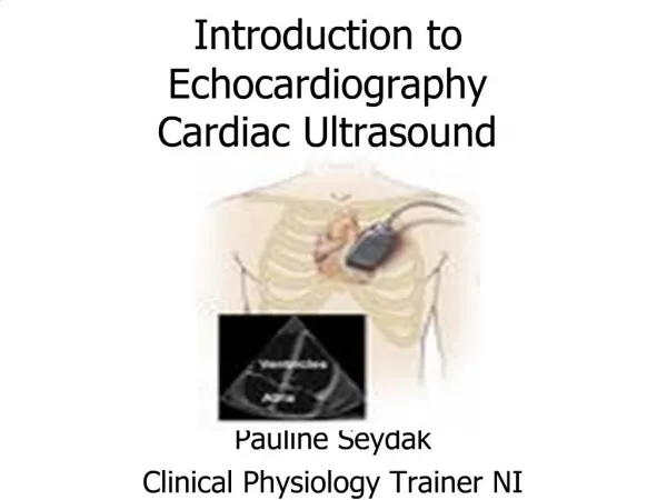
Introduction to Ultrasound VCA 341 Meghan Woodland, DVM March 16, 2012.
Indications • As a compliment to abdominal radiographs • To rule in/out intestinal obstruction (foreign body) • To determine the origin of an abdominal mass • Spleen, Liver • To facilitate fine needle aspiration/cystocentesis • To evaluate organ parenchyma • To assess fetal viability in pregnant animals • ***If clinical signs or history indicate abdominal ultrasound, then it should be performed even if radiographs are normal!!!
Pitfalls of Ultrasound • Ultrasound cannot penetrate air or bone • May be difficult to assess the GI tract in animals with aerophagia • Size of organs is largely subjective • Except renal size in cats • Unable to evaluate extra-abdominal structures • May still need to perform abdominal radiographs • Cost • User dependent results
Why do you need both? • Examples • Prostatic adenocarcinoma seen on ultrasound • Has it spread to the lumbar vertebrae? • Coughing patient with mitral regurgitation on echocardiogram • Does the patient have pulmonary edema? • Enlarged liver on radiographs • Can get a guided FNA with ultrasound
Examples • Prostate Abnormal Normal (Neutered Dog)
Need radiographs to properly evaluate the spine for metastasis
Ultrasound Physics • Characterized by sound waves of high frequency • Higher than the range of human hearing • Sound waves are measured in Hertz (Hz) • Diagnostic U/S = 1-20 MHz • Sound waves are produced by a transducer
Ultrasound Physics • Transducer (AKA: probe) • Piezoelectric crystal • Emit sound after electric charge applied • Sound reflected from patient • Returning echo is converted to electric signal grayscale image on monitor • Echo may be reflected, transmitted or refracted • Transmit 1% and receive 99% of the time
Attenuation • Absorption = energy is captured by the tissue then converted to heat • Reflection = occurs at interfaces between tissues of different acoustic properties • Scattering = beam hits irregular interface – beam gets scattered
Acoustic Impedance • The product of the tissue’s density and the sound velocity within the tissue • Amplitude of returning echo is proportional to the difference in acoustic impedance between the two tissues • Velocities: • Soft tissues = 1400-1600m/sec • Bone = 4080 • Air = 330 • Thus, when an ultrasound beam encounters two regions of very different acoustic impedances, the beam is reflected or absorbed • Cannot penetrate • Example: soft tissue – bone interface
Frequency and Resolution • As frequency increases, resolution improves • As frequency increases, depth of penetration decreases • Use higher frequency transducers to image more superficial structures • Ex: Equine Tendons Frequency Penetration
Instrumentation - Ultrasound Probes A B C A B C
Transducers/Probes • Sector scanner • Fan-shaped beam • Small surface required for contact • Cardiac imaging • Linear scanner • Rectanglular beam • Large contact area required • Curvi-linear scanner • Smaller scan head • Wider field of view
Monitor and Computer • Converts signal to an image/ archive • Tools for image manipulation • Gain – amplification of returning echoes • Overall brightness • Time gain compensation (curve) • Adjust brightness at different depths • Freeze • Depth • Zoom in for superficial view • Zoom out for wide view • Depth limited by frequency • Focal zone • Optimal resolution wherever focal zone is
Modes of Display • A mode • Spikes – where precise length and depth measurements are needed – ophtho • B mode (brightness) – used most often • 2 D reconstruction of the image slice • M mode – motion mode • Moving 1D image – cardiac mainly
Artifacts • Artifacts lead to the improper display of the structures to be imaged • Affect the quality of images • Improper machine settings – gain • Image too bright or too dark • Can disguise underlying pathology
Artifacts • Reverberation • Time delays due to travel of echoes when there are 2 or more reflectors in the sound path • Mirror image – liver, diaphragm and GB • Return of echoes to transducer takes longer because reflected from diaphragm • A second image of the structure is placed deeper than it really is • Comet tail – gas bubble • Ring down – skin transducer surface
Mirror Image Artifact Dr. Matthews
Artifacts • Acoustic shadowing • U/S beam does not pass through an object because of reflection or absorption • Black area beyond the surface of the reflector • Examples: cystic calculi, bones • Acoustic enhancement • Hyperintense (bright) regions below objects of low U/S beam attenuation • AKA Through transmission • Examples: cyst or urinary bladder
Artifacts • Refraction: • Occurs when the sound wave reaches two tissues of differing acoustic impedances • U/S beam reaching the second tissue changes direction • May cause an organ to be improperly displayed
Ultrasound Terminology • Never use dense, opaque, lucent • Anechoic • No returning echoes= black (acellular fluid) • Echogenic • Regarding fluid--some shade of grey d/t returning echoes • Relative terms • Comparison to normal echogenicity of the same organ or other structure • Hypoechoic, isoechoic, hyperechoic • Spleen should be hyperechoic to liver • Liver is hyperechoic to kidneys
Patient Positioning and Preparation • Dorsal recumbency • Lateral recumbency • Standing • Clip hair • Be sure to check with owners • Apply ultrasound gel • Alcohol can be used – esp. in horses
Image Orientation and Labeling • Must be consistent • Symbol on screen ~ dot on transducer • “dot” to head and “dot” to patients right • “dot” lateral for transverse and proximal for longitudinal images • Label images carefully • Organ • Patient’s name • Date of examination
Ultrasound-Guided FNA/ Biopsies • NORMAL ABD U/S FINDINGS DO NOT MEAN ORGANS ARE NORMAL!!! • ***Do FNA if suspect disease • Abnormal U/S findings nonspecific • Benign and malignant masses identical • Bright liver may be secondary to Cushing’s dz or lymphoma • Aspirate abnormal structures (with few exceptions)!!! • Obtain owner approval prior to exam • Warn owner of risks • +/- Clotting profile
Ultrasound-Guided FNA/ Biopsies • Risks of FNA’s • Fatal hemorrhage • Pneumothorax w/ pulmonary masses • Seeding of tumors • TCC • Sepsis • Abscesses
Ultrasound-Guided FNA/ Biopsies • Routinely aspirate: • Liver (masses and diffuse disease) • Spleen (nodules and diffuse disease) • Gastrointestinal masses • Enlarged lymph nodes • Enlarged prostate • Pulmonary/ mediastinal masses (usually don’t biopsy due to risk of pneumothorax • Occasionally aspirate: • Kidneys (esp. if enlarged) • Pancreas • Urinary bladder masses • Never aspirate: • Adrenal glands • Gall bladder
Ultrasound-Guided FNA/Biopsies • Non-aspiration Technique • 22g 1.5in needle • 6 cc syringe • Short jabs into organ • Spray onto slide, smear, and check abdomen for hemorrhage
Ultrasound-Guided FNA • Aspiration technique • Same set up as with non-aspiration technique • With needle in structure, pull back plunger vigorously several times • Remove needle, fill syringe with air • Spray onto slide and smear
Ultrasound-Guided Core Biopsies • Use a special biopsy “gun” • 14-20g • Insert through small skin incision • Much more representative sample • Tissue not just cells • Sometimes it is necessary to get the answer • But…. MUCH MORE LIKELY TO BLEED!
Summary • Know your limitations • Lack of expertise • $15,000 vs. $150,000 machine • For abdomen or thorax, do radiographs first • If safe and reasonable, do FNA’s of all suspected abnormal structures based on history, clinical signs, or the ultrasound examination • Abnormal structures can look normal • Of the structures that do look abnormal, benign and malignant processes can be identical • Documentation – save images in some fashion