Planmeca Intra
Planmeca Intra. Bisecting angle technique. Patient holds the film or sensor with his finger Short cone magnified apexes. Parallel technique. Film/sensor holder Long cone. Image quality. Small focal spot 0.7mm -> small unsharpness Optional long cone small unsharpness
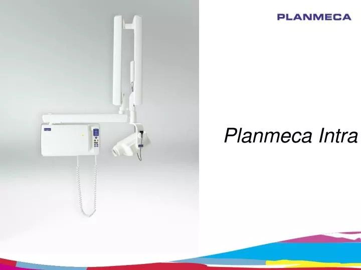

Planmeca Intra
E N D
Presentation Transcript
Bisecting angle technique • Patient holds the film or sensor with his finger • Short cone • magnified apexes
Parallel technique • Film/sensor holder • Long cone
Image quality • Small focal spot 0.7mm -> small unsharpness • Optional long cone • small unsharpness • Geometric requirements for good images are met
Image quality • High-voltage generator • Constant potential (DC) X-ray generation • High operating frequency (66 kHz) • 25% less radiation to patient compared to conventional AC generators • Shorter exposure times • Improved contrast • Is not affected by line voltage variations • Readiness for digital systems
Kilovolts and image quality Lower anode voltage • higher contrast, more suitable for endodontic, apex and bone structure diagnosis Medium anode voltages • boarder grey scale, suitable for caries detection Higher anode voltages • longest grey scale spectrum for periodontal disease diagnosis 50 kV 60 kV 70 kV
Milliampers and image quality With variable milliamperes (2 - 8 mA) we can take the whole advantage of the modern digital imagingsystems and new high-speed films.
Adjustable settings • adjustable kV setting (50, 53, 55, 57, 60 ,63, 66, 70) • different diagnostic needs are fulfilled • adjustable mA (2-8 mA) • maximum dose reduction possible with modern digital sensor systems and high-speed films
X-ray arm • smooth movements • drift-free positioning • no vibrations • easy, quick and accurate positioning
X-ray Tube design • non-symmetric form • tubehead and cone have a common smooth plane • easy targeting along the smooth surface • close to the patient’s chest in occlusal images
Controls 66 pre-programmed quick settings • modality selection to choose film, imaging plate or sensor • density setting • adult/child selection • periapicals for different teeth • occlusal • bite-wing / endo • Quick setting allow ALWAYS to have right exposure values for individual cases
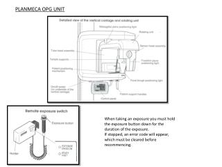
Control panel options • hand held control panel • remote exposure station • all controls easily at hand for exposure
Standard wall-mount • 4 extension arm lengths • reach 1525 – 1975 mm • special lengths available with custom order up to 2300 mm
Other mounting alternatives • dental unit mount • mobile base mount
Other mounting alternatives • ceiling mount • ceiling mount with operating light • floor column mount • single stud mount • pass-through mount
The End More information: Erkki Hiltunen Product Manager, X-rays tel: +358 20 7795 456 erkki.hiltunen@planmeca.com Mark Niemi Product Manager, X-rays tel: +358 20 7795 743 mark.niemi@planmeca.com More information: Osku Sundqvist Product Manager, Software tel: +358 20 7795 793 osku.sundqvist@planmeca.com 4/2011