Download
1 / 56
560 likes | 750 Vues
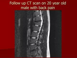
CT Radiographic Unknowns 66 yr old female with back pain. RADIOGRAPHIC FILM REPORT Radiographic Procedure: CT Scan Lumbar Spine Clinical Information: A 66 year old female with back pain

E N D
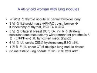
CT Radiographic Unknowns 66 yr old female with back pain
RADIOGRAPHIC FILM REPORT Radiographic Procedure: CT Scan Lumbar Spine Clinical Information: A 66 year old female with back pain Radiographic Findings: Lateral lumbar spine film shows loss of height at the T-12 vertebral body of 25% with end plate buckling. Axial CT scans at bone windows with reconstructed saggital images show compression deformity at T-12 with some anterior wedging. There is retropulsion of fragments into the spinal canal best seen on reconstructed views. This is causing moderate compromise of the canal of approximately 50%. Conclusion: Vertebral body fracture with 25% loss of height at T-12. This shows no obvious lytic component to suggest a metastatic lesion. There is some compromise of the central canal. Correlation with symptoms of cord or root compression is suggested.
CT Radiographic Unknown 60 yr old with screening physical
RADIOGRAPHIC FILM REPORT Radiographic Procedure: CT Scan Thorax Clinical Information: A 60 year old male with abnormal CXR on screening physical Radiographic Findings: Frontal chest radiograph shows shallow inspiration. Dual lead pacemaker is in normal position. The cardiac silhouette is normal. There is increased soft tissue density extending into the superior mediastium distorting norml anatomy. Impression: Abnormal CXR. The possibility of adenopathy or mass would have to be considered. CT scan is suggested if no old films are available to indicate this as a static process. Axial CT scans of the chest show a single section at the level of the aortic arch. IV contrast is seen in the SVC and aortic arch. There is low density material seen extending at the margins of the arch anteriorly. This tissue is in a fat density range. Conclusion: Mediastinal fat at the level of arch accounts for findings on CT scan. This indicates a benign process, mediastinal lipomatosis. This can be seen in patients receiving steroid treatment.
NUCLEAR MEDICINE TOMOGRAPHY S ingle P hoton E mission C omputed T omography
NUCLEAR MEDICINE TOMOGRAPHY P ositron E mission T omography
Sagittal Trans-axial Normal perfusion pattern
CHARACTERISTICS OF AN IDEALRADIOPHARMACEUTICAL 1. Pure gamma ray emitter 2. Gamma energy between 100 KeV and 200 KeV 3. Short physical half life 4. Readily available 5. Good chemistry
TYPES OF HALF-LIVES 1. Physical half-life 2. Biological half-life 3. Effective half-live
EFFECTIVE HALF LIFE T 1/2 Biologic x T 1/2 Physical = T 1/2 Effective T 1/2 Biologic + T 1/2 Physical
Anterior cerebrals Right middle cerebral division Right carotid Rt. Lt. “Stroke”
Posterior Anterior Rt. Lateral Lt. Lateral
Sagittal Trans-axial Normal perfusion pattern
Sagittal Trans-axial Normal perfusion pattern
Lt. SSN Normal Technetium 99m image
Anterior Posterior
Anterior Posterior
Perfusion Ventilation Matched defects indicate intrinsic lung disease
Normal ventilation and abnormal perfusion indicates pulmonary embolism
Anterior Normal Liver
Rt. Lateral Anterior Space occupying lesions
Early Late Biliary Tract Study
Normal Abnormal Myocardial Perfusion Study
Good function Bad function Myocardial Function Test
Normal Abnormal Infarct Imaging
Radiographic Film Report Radiographic Procedure: Abdomen – flat film Clinical Information:LUQ mass Radiographic findings: An AP, flat film of the abdomen showed splenomegaly. The vertical dimension of the spleen was approximately 17cm. The liver was at the upper limits of normal size. The bowel gas pattern was normal. The GI tube was looped in the stomach although the tip had advanced into the second portion (descending) of the duodenum. Conclusion: There is splenomegaly. Lumbar scoliosis was noted.